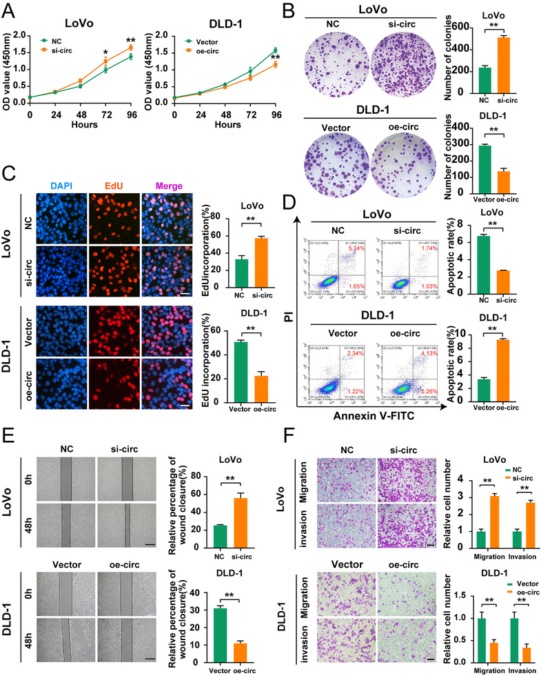
FIGURE 2.

Hsa_circ_0001666 suppresses the proliferation, invasion and induces the apoptosis of CRC cells in vitro. LoVo cells were transfected with si‐circ or NC, and DLD‐1 cells were transfected with oe‐circ or Vector. (A–C) To evaluate the proliferative ability, CCK‐8 assays, colony formation assays and EdU assays were used (magnification, 200×; scale bar, 100 μm). (D) The Annexin‐V FITC/PI staining was used to assess apoptotic rates. (E. F) Wound healing assays and transwell assays were used to evaluate the migratory and invasive capabilities. The scale bar in wound healing assays indicated 20 μm; the scale bar in transwell assays indicated 200 μm. Data were all represented as mean ± SD (n = 3). *p < .05, **p < .01
