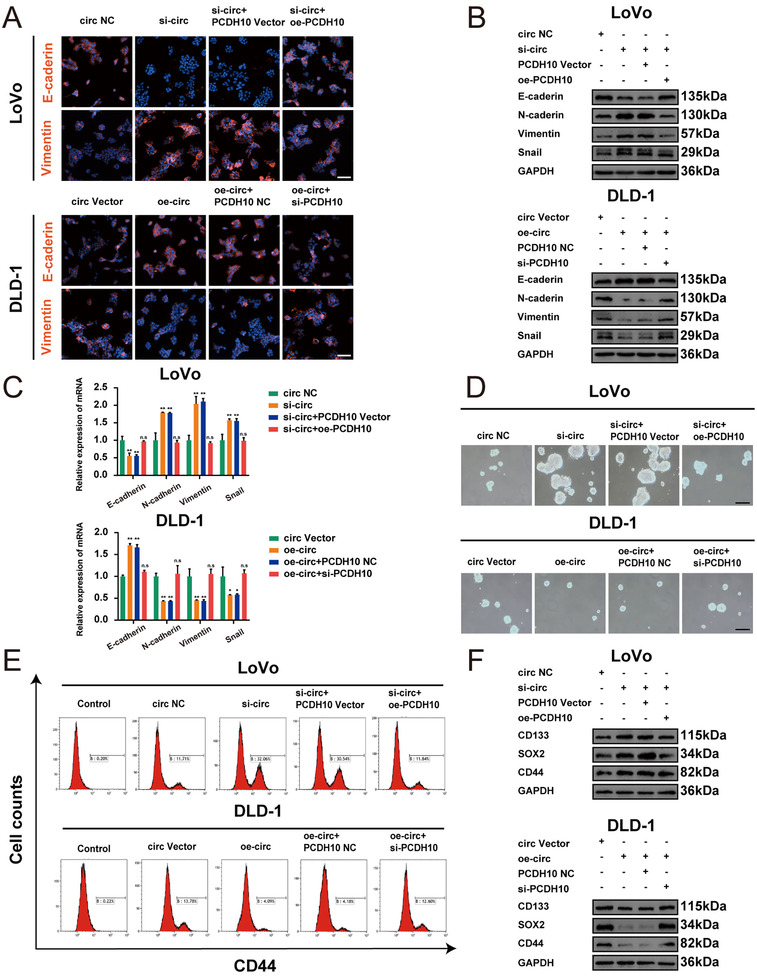
FIGURE 8.

Hsa_circ_0001666 suppresses EMT and cell stemness by miR‐576‐5p/PCDH10 axis in vitro. LoVo cells were transfected with circ NC, si‐circ, si‐circ+PCDH10 Vector, si‐circ+oe‐PCDH10, and DLD‐1 cells were transfected with circ Vector, oe‐circ, oe‐circ+PCDH10 NC, oe‐circ+si‐PCDH10. (A) The expression of E‐cadherin and Vimentin were detected using IF; scale bars, 50 μm. (B,C) The expression of EMT marker genes was detected by Western blot and qRT‐PCR. (D) Typical images from the sphere formation assay. (E) The number of cells with the CD44+ phenotype. (F) The expression of stemness marker genes was detected by Western blot. Data were represented as mean ± SD (n = 3); n.s indicated no significance, **p < .01
