FIGURE 1.

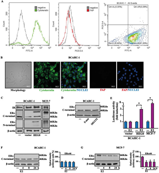
Molecular features of BCAHC‐1 cells. (A) Expression of cytokeratin and fibroblast activation protein (FAP) in BCAHC‐1 cells, as shown by flow cytometry. Representative overlay histograms and dot plot showing cytokeratin‐positive (green) and FAP‐negative (red) BCAHC‐1 cells. FITC, fluorescein isothiocyanate; PE‐Cy5.5, phycoerythrin‐Cyanin 5.5. (B) Morphological appearance of BCAHC‐1 cells in phase‐contrast microscopy, cytokeratin‐positive (green signal), and FAP‐negative (red signal) immunofluorescence staining in BCAHC‐1 cells. Nuclei were stained by DAPI (blue signal). Scale bar: 100 μm. (C) Immunoblots of IR, ERα66, and ERα46 in BCAHC‐1 and MCF‐7 breast cancer cells, as indicated. BCAHC‐1 cells were transiently transfected with an empty vector (vector) or the wild‐type ERα (HEG0). (D) Protein levels of ERα46 in BCAHC‐1 cells exposed for 24 h to 10 μM DNA‐hypomethylating agent 5‐aza‐2′‐deoxycytidine (5‐aza) and proteasome inhibitor MG132. β‐Actin was used as a loading control. (E) BCAHC‐1 and MCF‐7 cells were transfected with the ER luciferase reporter plasmid ERE‐luc combined with an empty vector (vector) or the wild‐type ERα (HEG0) for 8 h and treated with 100 nM E2 for 18 h, as indicated. The luciferase activities were normalized to the internal transfection control, and values of cells receiving vehicle (−) were set as 1‐fold induction on which the activity induced by E2 was calculated. Columns represent the mean ± SD of three independent experiments performed in triplicate. Protein levels of ERα46 in BCAHC‐1 (F) and ERα66 in MCF‐7 (G) cells treated with vehicle (−) or 100 nM E2 for the indicated times. Side panels show densitometric analysis of the blots normalized to β‐actin. Values represent the mean ± SD of three independent experiments performed in triplicate. (*) indicates p < 0.05 for cells treated with E2 relative to cells treated with vehicle (−)
Abbreviations: E2, 17β‐estradiol; ERα46, 46 kDa ERα splice variant; ERα66, 66 kDa estrogen receptor α; FAP, fibroblast activation protein; IR, insulin receptor; SD, standard deviation.
