FIGURE 4.

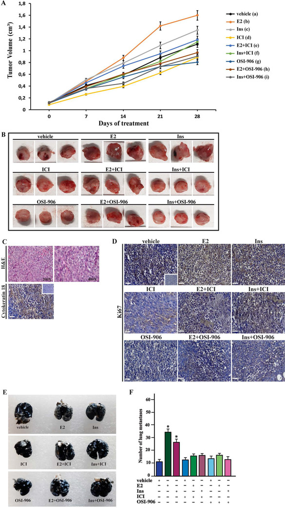
E2 and insulin induce the growth of BCAHC‐1‐derived xenografts and lung metastases. (A) Tumor volume from BCAHC‐1 xenografts implanted in female athymic nude mice treated for 28 days with vehicle, E2, insulin (Ins), ICI 182,780 (ICI), OSI‐906 alone or in combination (see details in Supplementary Materials and Methods). Tumor growth was monitored by caliper measuring the visible tumor sizes at indicated time points. Vehicle (a); E2 (b); Ins (c); ICI (d); OSI‐906 (e); E2+ICI (f); E2+OSI‐906 (g); Ins+ICI (h); Ins+OSI‐906 (g). p < 0.05 at the end of experiment among the following groups: b versus a; c versus a; d versus a; e versus a; f versus b; g versus b; h versus c; i versus c. (B) At the end of experiment, tumors were explanted, and for each group of animals (n = 6), three representative images of tumors are shown. (C) Formalin‐fixed paraffin‐embedded (FFPE) sections of tumor xenografts were stained with hematoxylin and eosin Y (H&E), and the epithelial nature of the tumors was verified by immunostaining with anti‐human cytokeratin 18 antibody. Scale bar: 25 μm. Insert: negative control. (D) The expression of Ki‐67, as a marker of proliferation, was evaluated in FFPE sections of explanted tumors from BCAHC‐1 xenografts treated as described in Supplementary Materials and Methods. Scale bar: 25 μm. Insert: negative control. (E) Representative photographs of lung metastases (white dots) after 15 days of treatment with vehicle, E2, insulin (Ins), ICI 182,780 (ICI), OSI‐906 alone or in combination (see Supplementary Materials and Methods). Lungs of xenografts were injected with India ink solution (15%) and fixed in Fekete's solution. (F) Enumeration of pulmonary metastatic nodules. The results are presented as mean ± SD (n = 6 lungs). (*) indicates p < 0.05 for animals treated with E2 and insulin (Ins) versus animals treated with vehicle
Abbreviations: E2, 17β‐estradiol; FFPE, formalin‐fixed paraffin‐embedded; SD, standard deviation.
