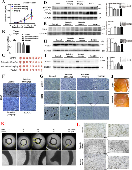
FIGURE 3.

Baicalein inhibits CRC growth and metastasis via TLR4/HIF‐1α/VEGF axis in vivo. (A) Tumor volume, (B–C) tumor weight of the CRC‐bearing xenograft mouse models after baicalein treatment. Western blot showing the expressions of (D) p‐NF‐κB, NF‐κB, (E) TLR4, (H) VEGF, and (I) MMP‐2 in the xenograft tissues. Immunohistochemistry staining of (F) Ki67, (G) VEGF, and CD31 in the xenograft tissues. (J) Blood vessel formation on the chick yolk sac membrane after baicalein treatment. Vessel sprouting in (K) the rat aortic ring model of angiogenesis, and (L) tube formation of the cultured human endothelial cells after baicalein treatment. Shown is mean ± SE, n = 8 mice in each group or n = 3 individual experiments, *P < 0.05, **P < 0.01 compared to control. VEGF, vascular endothelial growth factor; MMP2, matrix metalloproteinases 2
