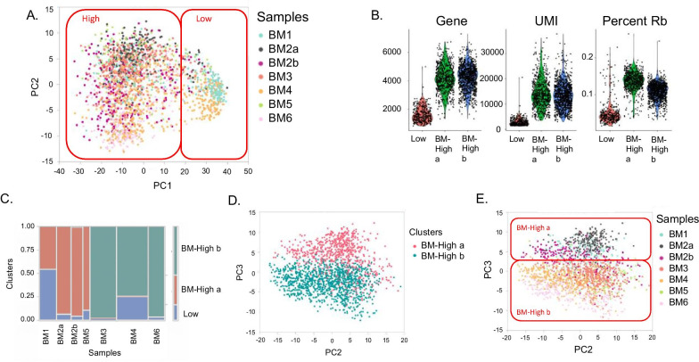
Fig. 1.
Clusters of BM-MSC transcriptome profiles. A The first two Principal Components of gene expression identify two broad clusters of cells, which are colored by sample: Cluster BM-Low, which corresponds to low UMI count cells and cluster BM-High, with high UMI count cells. B Violin plots show the density of the number of Genes, UMI, and Ribosomal Protein transcripts (RP) per cell. C Association of cells with clusters. The width of each column is proportional to the number of cells in the indicated [47] sample, and the color of each box corresponds to cells in cluster BM-Low (blue), BM-High_a (red) or BM-High_b (green). D Within cluster High, SC3 identifies two clusters of cells, which separate along PC3 as indicated by the red and blue points. E Shading of cells by sample confirms that cells from each donor belong to one of the two sub-clusters, although with subtle separation associated with PC3

