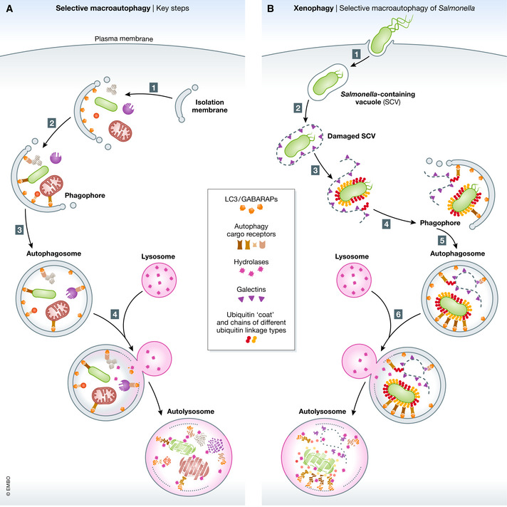
Figure 1. Selective macroautophagy.

(A) Key steps in selective macroautophagy. (1) The process of macroautophagy is initiated by isolation membranes and vesicles that gradually expand and mature to phagophores decorated with membrane‐anchored LC3 and GABARAPs (orange hexagons). (2) These act as a binding site for autophagy cargo receptors (brown) allowing direct delivery and accumulation of cellular cargo. Finally, the phagophore closes forming a double‐membraned vesicle called autophagosome (3), which subsequently fuses with the lysosome forming an autolysosome (4), where the content is degraded by lysosomal enzymes. (B) Xenophagy, selective macroautophagy of Salmonella. (1) Salmonella enters host cells by forming a Salmonella‐containing vacuole (SCV) that protects it from the host surveillance system and serves as a replicative niche. (2) SCVs can rupture allowing access of host cytosolic proteins. (3) The exposed glycans, that are normally present on the outer side of the cell membrane, serve as a danger signals that are recognized by galectins (purple triangles). Besides, Salmonella is detected by E3 ligases that generate a dense Ub coat, consistent of different linkage‐type Ub chains (red and orange circles), around Salmonella. (4) Both, galectins and Ub recruit a variety of autophagy receptors (brown) that mediate Salmonella capture by LC3‐conjugated phagophores and Salmonella‐containing autophagosomes (5) are targeted for lysosomal degradation (6).
