
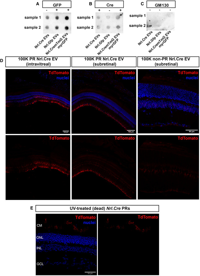
Figure EV1. Photoreceptor‐derived EV subpopulations contain Cre and GFP protein.
-
A–CRepresentative dot blots of GFP (A), Cre (B) and GM130 (C) expression in P8 Nrl.Cre+/− , Nrl.Gfp+/+ , Nrl.Cre+/− × mTmG +/+ (myrGFP) photoreceptor‐derived 100 K EV pellets, as appropriate (N = 8 experiments, each dot represents a pool from three independent EV isolations, derived from 60*106 cells from two samples). The lack of GM130 staining confirms the absence of contamination from Golgi within the EV preparations. Positive GM130 staining from whole cell lysate is shown in Fig 1.
-
DRepresentative tile‐scan images following (left) subretinal and (middle) intravitreal injection of EVs (100 K fraction) derived from P8 Nrl.Cre+/− photoreceptors, compared with (right) subretinal injection of EVs (100 K fraction) derived from non‐photoreceptors (Nrl.Cre+/− ); red = TdTomato recombined cells; blue = nuclei; Scale bar = 100 µm (left & middle) and 50 µm (right).