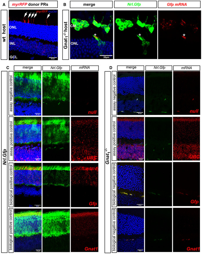
Figure EV4. Transplantation of Nrl.Gfp+/+ photoreceptors results in the transfer of cytoplasmic protein and some membrane‐bound protein, but little or no mRNA.

-
ARepresentative MIP confocal images of wild‐type (wt) eye cups fixed 21 days post‐transplantation with P8 myrRFP+ve CD73+ve MACS‐enriched photoreceptors. Note the presence of RFP+ host inner segments (arrows). myr‐RFP (red), nuclei (blue).
-
BRepresentative MIP confocal images of Gnat1 −/− eye cups fixed and stained in situ (RNAScope) with Gfp mRNA probe (red) 21 days post‐transplantation with P8 Nrl.Gfp+/+ photoreceptors (green). Blue = Dapi (nuclei); CM: cell mass; ONL: Outer Nuclear Layer. Asterisks denote a rare example of an integrated donor photoreceptor located within the host ONL and presenting robust staining for Gnat1 mRNA in the inner segment.
-
C, DRepresentative MIP confocal images of (C), Nrl.Gfp+/+ and (D), Gnat1 −/− eye cups fixed and stained in situ (RNAScope) with a null probe (assay negative control, red), UBC mRNA probe (assay positive control, red), Gfp mRNA probe (biological positive control, red), Gnat1 mRNA probe (biological positive control, red), and Gnat1 −/− tissue stained with either Gfp mRNA probe (biological negative control, red) or Gnat1 mRNA probe (biological negative control, red). Nrl.Gfp+/+ photoreceptors (green), nuclei (blue); mRNA (red).
Data information: Scale bars = 20 µm.
