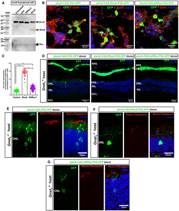
Figure EV5. Manipulation of actin dynamics by overexpression of RhoA or DNRac1 in transplanted photoreceptors correlates with reduced GFP and rod α‐transducin transfer.

-
ARepresentative Western blots of RhoA and Rac1 expression in mixed P0–2 retinal cultures transduced with plenti‐CAG‐P2A.GFP, plenti‐CAG‐ΔΝRac1‐P2A.GFP or plenti‐CAG‐RhoA‐P2A.GFP compared to β‐actin and assessed after 8 DIC.
-
BRepresentative MIP confocal images of P0–2 retinal cultures transduced with plenti‐CAG‐P2A.GFP (green) or plenti‐CAG‐RhoA‐P2A.GFP (green) or plenti‐CAG‐ΔΝRac1‐P2A.GFP (green), fixed and stained with F‐actin (red) (N = 2 independent cultures, n = 4 wells per condition); Scale bars = 20 µm.
-
CQuantification and statistical analysis of the effect of overexpression of RhoA and ΔΝRac1, versus control, on actin in P0–2 retinal cultures at 7DIC. Effect assessed by measurement of mean intensities (Image J) of F‐actin plaques (red) in GFP+ cells (green), normalized to GFP+ cell number; control 2.6 ± 0.7; RhoA 13.9 ± 2.1, ΔΝRac 4.5 ± 1.3. Mean ± SD. One‐way ANOVA non‐parametric two‐tail, Kruskal–Wallis post‐test ***P < 0.001 (N = 2 independent cultures, n = 4 wells per condition).
-
DRepresentative tile‐scan images of Gnat1 −/− eyes transplanted with P2 retinal cells transduced with plenti‐CAG‐P2A.GFP (green) or plenti‐CAG‐RhoA‐P2A.GFP (green) or plenti‐CAG‐ΔΝRac1‐P2A.GFP (green). Green = GFP, blue = Dapi (nuclei); Scale bars = 50 µm.
-
E–GRepresentative MIP images of Gnat1 −/− eyes transplanted with P2 retinal cells transduced with (E), plenti‐CAG‐P2A.GFP (green) or (F), plenti‐CAG‐RhoA‐P2A.GFP (green) or (G), plenti‐CAG‐ΔΝRac1‐P2A.GFP (green). Immunostaining for Rod α‐transducin indicates that inhibition of actin polymerization impairs transfer of Rod α‐transducin alongside that of GFP. Green = GFP, blue = Dapi (nuclei), red = Rod α‐transducin; Scale bars = 50 µm.
Data information: RPE = retinal pigment epithelium, CM = cell mass, ONL = outer nuclear layer, INL = inner nuclear layer, GCL = ganglion cell layer. All eyes were fixed and examined 21 days post‐transplantation.
