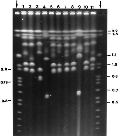
FIG. 1.
Representative electrophoretic karyotype patterns of C. parapsilosis. Each numbered lane corresponds to a cluster of overlapping karyotypes, as defined in the text. Arrowed lanes at left and right are S. cerevisiae chromosomal markers, and their molecular size is expressed in megabases. For other details, see the text.

