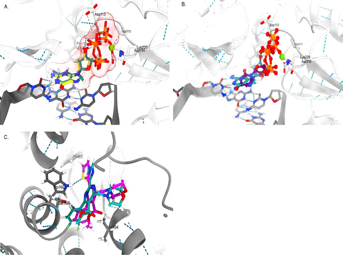
Figure 5.
3D representations of the (A) co-crystallized ligand 2-deoxyguanosine-5-triphosphate before (light sea green) and after docking (yellow) with HBV-Pol, (B) alignment of LAM (purple) compared to 2-deoxyguanosine-5-triphosphate (light sea green), and (C) HAP with HBV-Core before (light sea green) and after docking (magenta).

