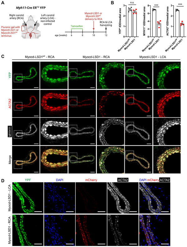
Figure 3: H3K4me2 editing impairs SMC contractile function in vivo.
A. Schematic of lentiviral-mediated H3K4me2 editing in vivo. B. Quantification YFP+, ACTA2+ and MYH11+ Integrated Optical Density (IOD) normalized to medial area in carotid cross-sections of Myh11-CreERT2 YFP mice locally infected with Myocd-LSD1 or Myocd-LSD1NF lentivirus (n=4 mice per group). C. YFP, ACTA2, and MYH11 staining in non-infected left carotid arteries (LCA), Myocd-LSD1 and Myocd-LSD1NF infected right carotid arteries (RCA). Scale bar = 100 μm. D. YFP, mCherry and Acta2 staining in non-infected left carotid or Myocd-LSD1 infected right carotid arteries. Scale bar = 100 μm. Data are represented as mean ± s.e.m of 4 independent biological replicates. Groups were compared by Student t-test (B). * p<0.05, ** p<0.001, *** p<0.0001.

