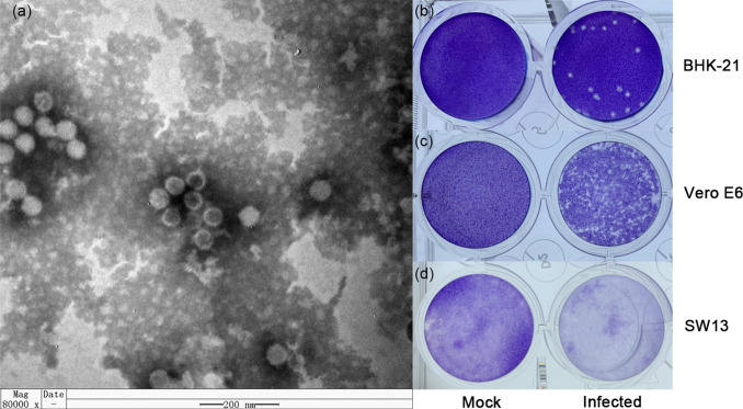
Fig. 1.
The viral morphology and viral plaques in cells of YN15-283-01. (a) Negative-stained ultracentrifuged virions. Viral plaques in (b) BHK-21 (10−5 dilution at four dpi), (c) Vero E6 (10−3 dilution at five dpi) and (d) SW13 (10−3 dilution at five dpi) cell monolayers. The wells were 16 mm in diameter.

