
Fig. 1.
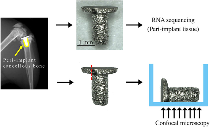
Peri‐implant tissue collection and immunofluorescence staining preparation. The implant was removed from the right tibias. (Yellow = cancellous bone around the implant.) One end of the tibial plateau component of the implant was polished before immunofluorescence staining.