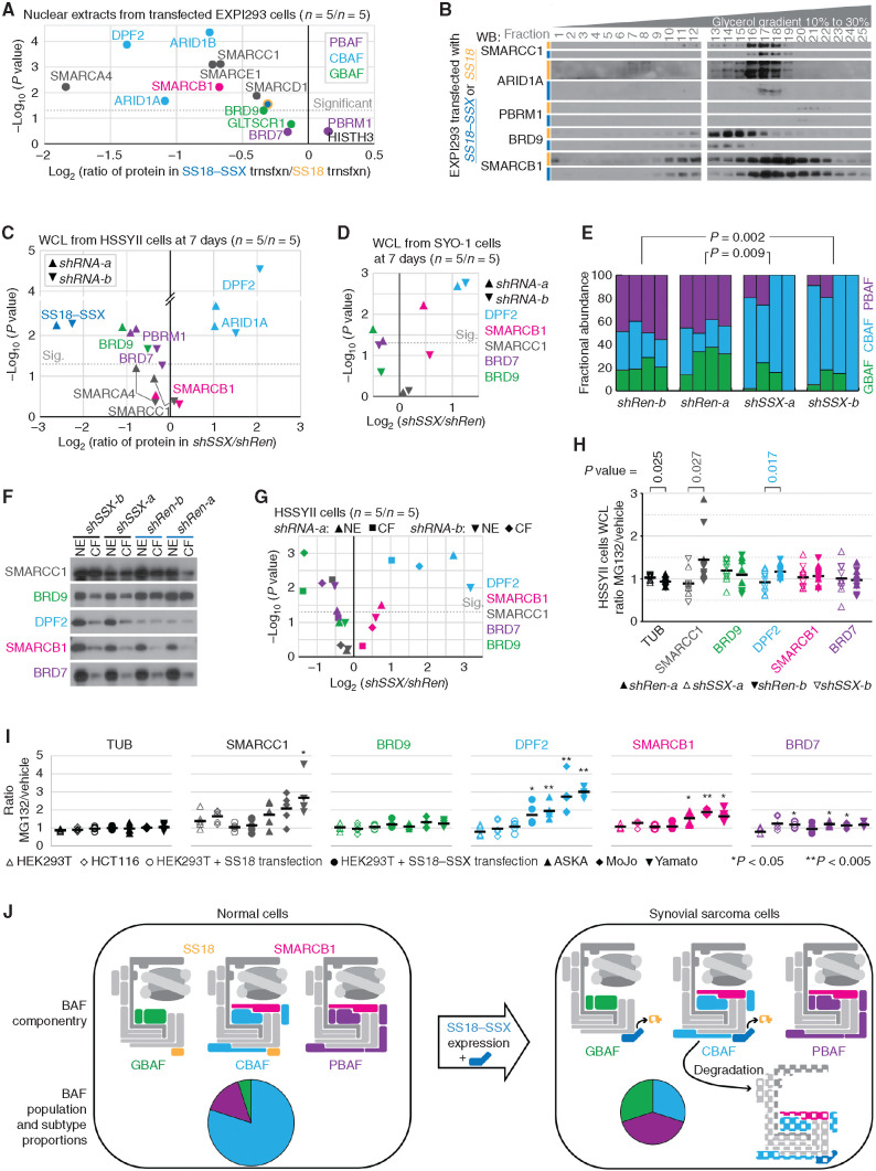
Figure 7.
Expression of SS18–SSX leads to CBAF complex reductions and relative overabundance of PBAFs and GBAFs. A, Quantitative LICOR WBs of BAF components in nuclear extracts of EXPI293 cells transfected with either SS18–SSX or SS18 (n = 5 each) presenting the log-transformed two-tailed Student t test P value of the difference between and the ratio of protein in the fusion-transfected versus SS18-transfected cells. trnsfxn, transfection. B, WBs of glycerol gradient fractions of EXPI293T cells transfected with SS18–SSX or SS18. C, LICOR quantitative WBs from whole-cell lysates (WCL) collected from HSSYII human synovial sarcoma cells subjected to 7 days of shRNAs (two sequences each; n = 5 for each sequence of each shRNA) directed against control (Renilla, shRen) or the fusion (SS18–SSX, shSSX), with BAF subunits color coded by BAF, presented as log-transformed paired two-tailed t test P values and ratios of fusion knockdown over control knockdown. Sig., significant. D, LICOR quantitative WBs of WCLs from SYO-1 human synovial sarcoma cells after knockdown of the fusion or control for 7 days. E, Fractional abundances of BAF subtypes defined by optical densitometry–quantified gradients of SMARCC1 (as in Fig. 6C) for HSSYII cells subjected to shRNAs against the fusion or control. (P values from two-tailed paired t tests; n = 4 for each shRNA.) F, WBs of nuclear extract (NE) with the paired chromatin fraction (CF; protein that stays with the insoluble chromatin pellet after NE) of proteins after 7 days of fusion or control knockdown. G, LICOR quantitative WB abundances presented as paired t test P values and ratios of fusion over control knockdown in each of the NE and CF components of HSSYII cells after 7 days. H, LICOR quantitative WB–defined proteins in WCLs presented as the ratios of MG132-treated over DMSO vehicle–treated cells after weeklong shRNA depletion of SS18–SSX or control Renilla (n = 5 for each condition, n = 10 for each group; two-tailed heteroscedastic t test comparing the ratios for each protein by knockdown group). I, LICOR quantitative WB–defined proteins in WCLs presented as of the ratios of MG132-treated over DMSO vehicle–treated cells from synovial sarcoma (or SS18–SSX-transfected) and control (or SS18-transfected) cell lines (n = 5 each; P values are from two-tailed Student t tests comparing each to HEK293T untransfected control cells). J, Model schematic of the impact of SS18–SSX expression on BAF componentry and relative abundance of BAF subtypes.

