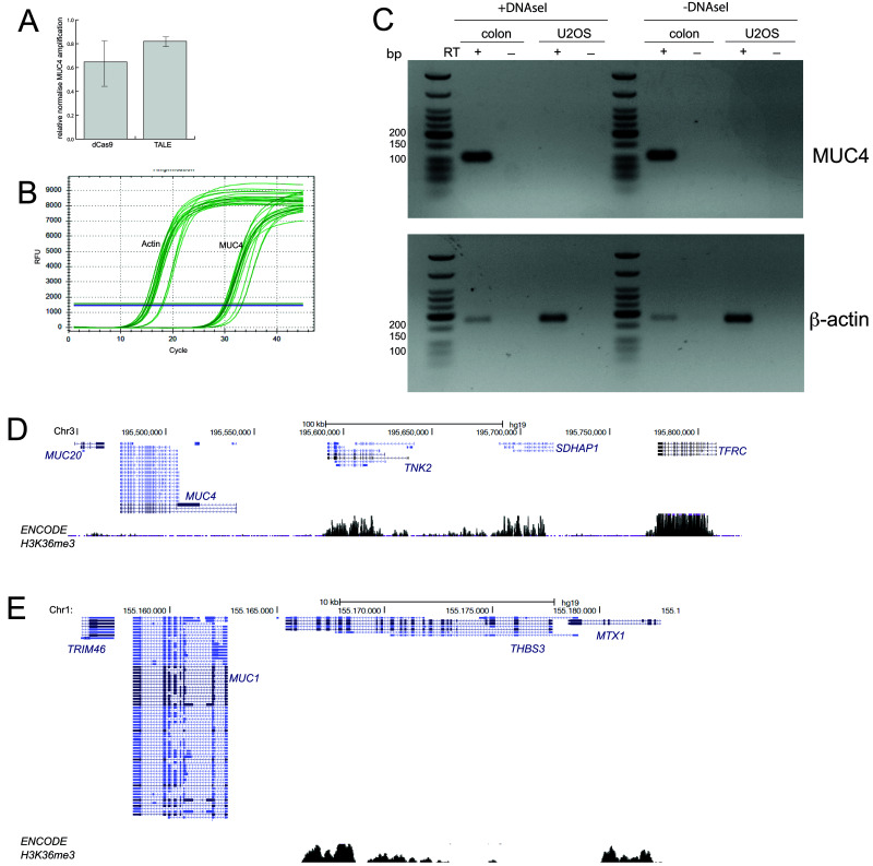
Figure 3. No evidence of MUC4 expression in U2OS cells.
A) qRT-PCR data for MUC4 showing amplification levels in cells transfected with dCas9 and TALEs targeting MUC4, relative to mock transfected cells (2 -ΔΔCt) and normalised to β-actin. Primers are those described by Chen et al. (2013) and shown in Figure 1B. Data show means (+/- stdev) from three biological replicates. B) Graph showing the rise in amplified product concentration with increasing cycle number for β-actin and for MUC4. Data shown are from three technical replicates of one of the three biological replicates used in ( A). C) RT-PCR using the primers shown in Figure 1C to detect the expression of MUC4 (top) in RNA prepared from U2OS cells and human colon mucosa. Amplification of β-actin (bottom) acts as a positive control. - reverse transcriptase (RT) and DNAse I untreated samples act as controls for the presence of genomic DNA contamination in RNA samples. D and E) UCSC Genome Browser screen shot of the genomic regions containing the MUC20/MUC4 ( D) and MUC1 ( E) loci and adjacent non-mucin genes. Shown below is the ENCODE H3K36me3 ChIP-seq track from U2OS cells (GEO Accession number GSM788076). Genome co-ordinates are from the hg19 assembly of the human genome.

