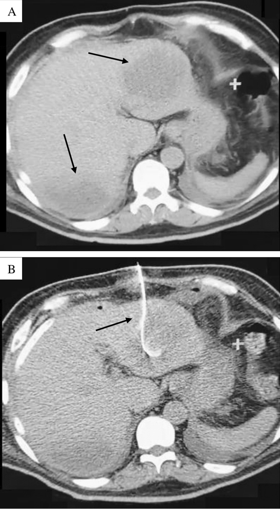
Fig. 2.

Computed tomography. A Axial section of abdominal computed tomography scan without contrast administration showing an enlarged liver (caudal skull diameter: 18 cm), at least two images were located in segments II and VIII of rounded morphology, regular edges, hypodense (Hounsfield Units: 20), the largest with 150 cc volume, likely related to liver abscess; B axial section of abdominal computed tomography scan without oral or intravenous contrast administration, showing the placement of a catheter for drainage of the liver abscess located in segment II
