Figure 3.
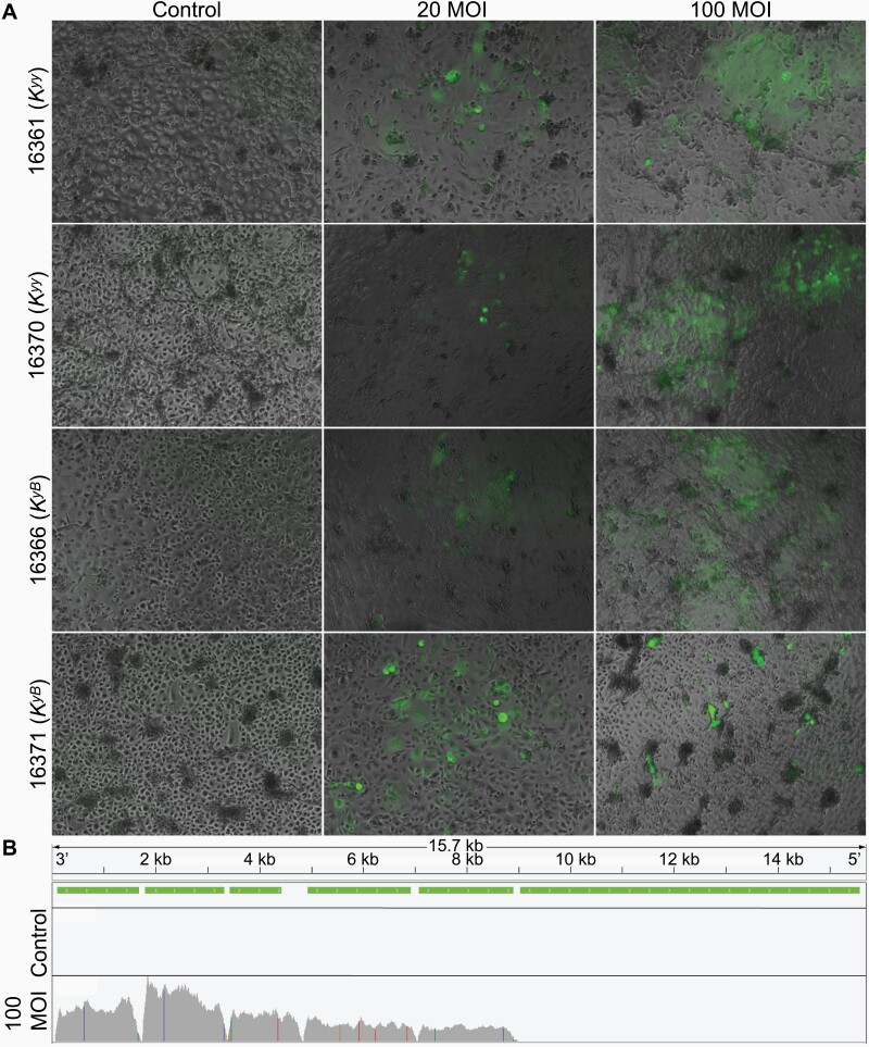
Primary wolf keratinocytes are vulnerable to canine distemper virus infection. (A) Kyy and KyB primary wolf keratinocytes infected with live canine distemper virus for 5 days. Keratinocytes were infected at an MOI of 20 and 100 TCID50/cell. Fluorescence images were captured to visualize CDV (expressing GFP) and overlaid on phase-contrast images. Animal IDs and corresponding CBD103 genotypes are indicated on the y axis. (B) Visualization of RNA-Seq read pile-ups (gray) across the full CDV genome from a representative wolf cell culture (animal ID 16361) treated for 5 days with CDV at 100 MOI or vehicle control. CDV transcription is evident from RNA extracted from infected cells, but not from control cells. Green bars indicate the genomic locations of CDV genes. Colored vertical lines indicate polymorphism relative to the reference genome.

