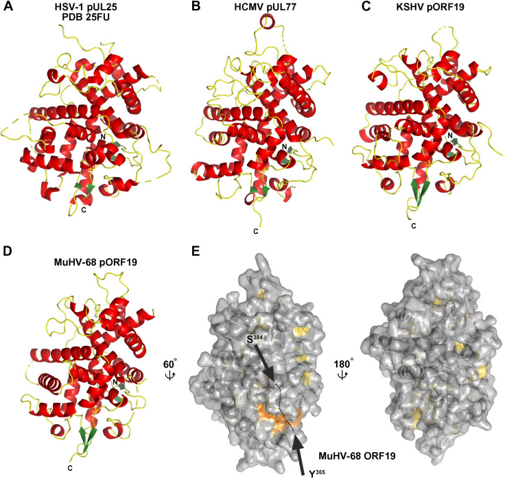
Fig 1. Crystal structure of the minor KSHV capsid protein pORF19 and its orthologs.
(A–D) Cartoon representation of CTDs derived from HSV-1 pUL25 (PDB 2F5U; 18), HCMV pUL77, KSHV pORF19, and MuHV-68 pORF19 colored according to secondary structure with α-helices in red, β-strands in green, and loop regions in yellow. Disordered regions are shown as dashed tubes, N-terminal and carboxyl terminus are indicated. (E) View on the MuHV-68 pORF19 CTD (pORF19MCTD) shown in surface representation with residues conserved across all 4 orthologs colored in yellow. A larger conserved patch consisting of 4 residues distant in primary sequence (colored in orange) is in close proximity to the loop connecting Y365 and S384 (black arrows), which is disordered in the crystal structures of all orthologs, suggesting that the conserved patch is likely buried in the native protein. CTD, carboxyl-terminal domain; HCMV, human cytomegalovirus; HSV, herpes simplex virus; KSHV, Kaposi’s sarcoma-associated herpesvirus; MuHV-68, murid gammaherpesvirus 68.

