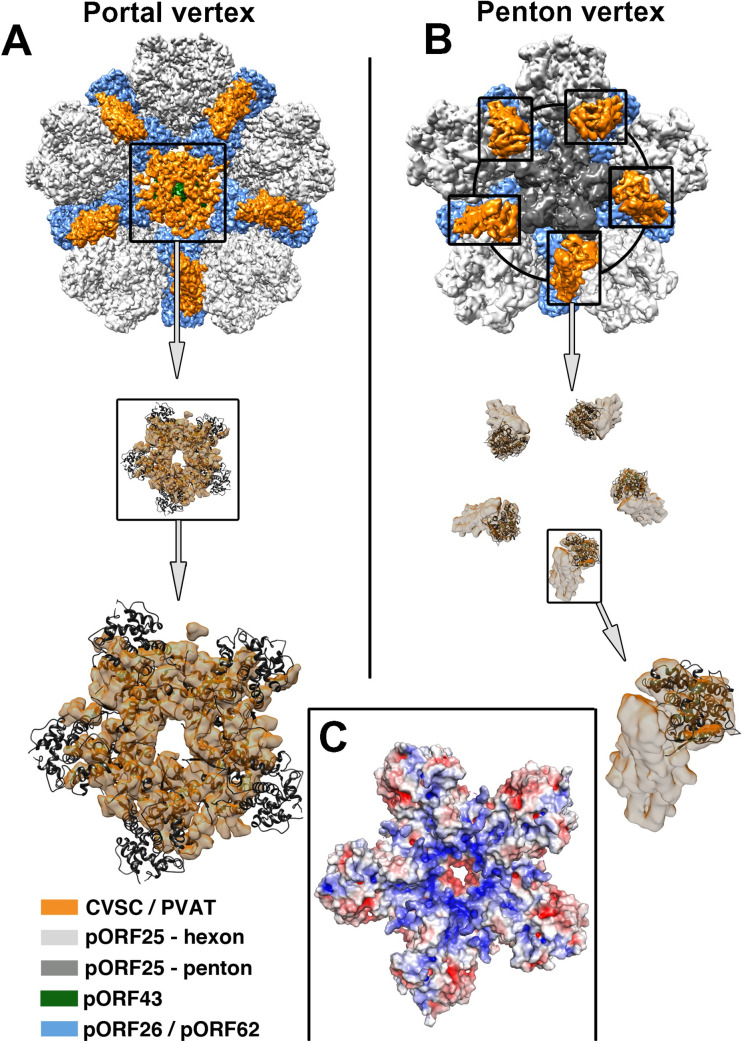
Fig 3. Arrangement of the pORF19KCTD pentamer on the capsid.
(A) Cartoon representation of the pentameric pORF19KCTD ring fitted into the portal cap electron density of the asymmetric (C1) KSHV portal vertex reconstruction (EMDB 20431; [4]) and (B) the pORF19KCTD protomers (colored in dark gray) fitted into the CVSC density of a symmetric KSHV penton vertex reconstruction (EMD 6038; [13]). Capsid proteins are colored as in S1 Fig. The close-up view (A, bottom panel) underlines the high quality of the fit (cross-correlation coefficient of 0.78 calculated by Chimera [40]). (C) The electrostatic potential on the molecular surface of the pentameric pORF19KCTD ring, represented ramp-colored from red (negative) to blue (positive) through white (neutral), calculated using APBS and contoured on a scale ranging from −5 to +5 kT/e, reveals an accumulation of positive charges in or around the funnel at the center of the pentamer, suggesting an electrostatic interaction with the phosphate backbone of the viral genome terminus. CVSC, capsid vertex–specific component; KSHV, Kaposi’s sarcoma-associated herpesvirus; PVAT, portal vertex–associated tegument.

