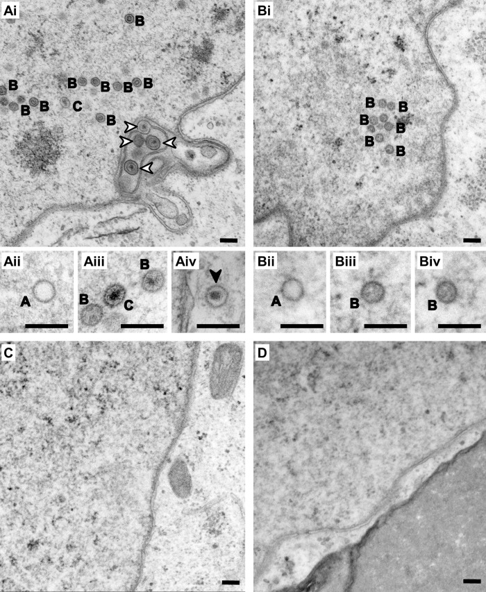
Fig 8. Formation of capsids in reactivated cells requires the presence of functional pORF19.
(A–D) Electron microscopy analysis of individual iSLK cell clones transfected with either wt or mutant KSHV-Bac16 constructs after KSHV reactivation. In cells transfected with wt KSHV-Bac16 (A) viral capsids were frequently found in the nucleus (Ai, Aii, and Aiii) and to a lesser extent in the cytoplasm (Aiv). In the nucleus, all stages of nuclear capsid assembly were identified, including A capsids (labeled with A), B capsids (labeled with B), and C capsids (labeled with C), as well as capsids in the process of primary envelopment at the nuclear envelope (Ai, white arrowheads). In cells transfected with KSHV-Bac16DQ (B), only nuclear capsids of the types A (labeled A in Bii) and B (labeled B in Biii and Biv), but no C capsids were observed. No capsids were found undergoing primary envelopment or in the cytoplasm. In cells transfected with KSHV-Bac16VL (C) or KSHV-Bac16loop (D), no viral structures were identified in the nucleus or the cytoplasm, similar to mock-transfected cells. Scale bars correspond to 200 nm. KSHV, Kaposi’s sarcoma-associated herpesvirus; wt, wild-type.

