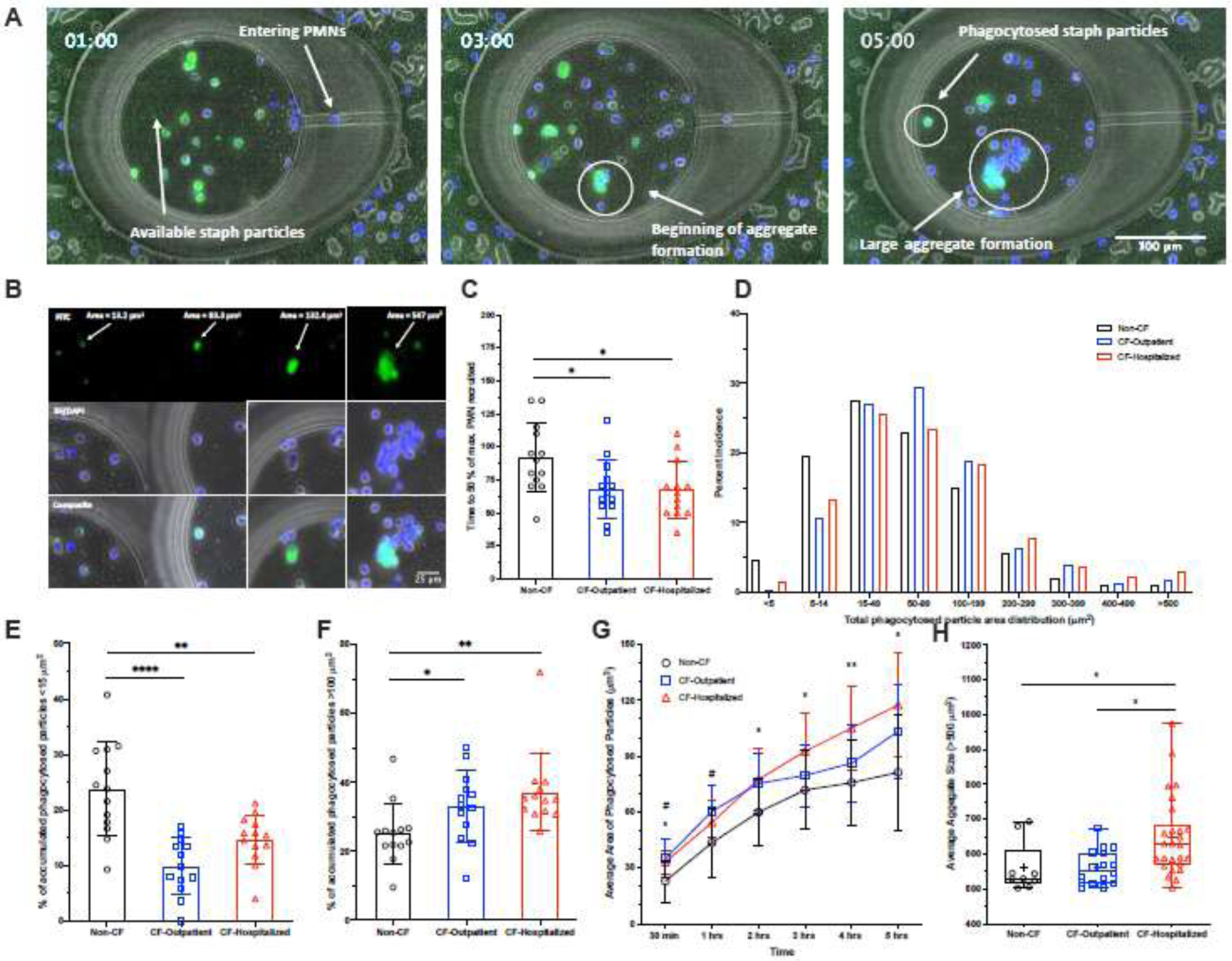
Figure 2: CF neutrophils display differences in microbe-like particle phagocytosis:

S. aureus bio-particles with fMLP were loaded into microfluidic chambers. Buffy coat-containing neutrophils stained with Hoechst were loaded around these chambers to observe host-pathogen interactions, specifically neutrophil recruitment to and phagocytosis of these particles. (A) A panel of images taken at 1, 3 and 5 hours showing the distribution of neutrophils (Hoechst, blue) and phagocytosis behaviors in response to S. aureus particles (FITC, green), which are a faint green, becoming brighter with increased aggregation. (B) A panel (left to right) showing small areas of phagocytosed particles formed by individual neutrophils to larger aggregates of S. aureus particles formed by multiple neutrophils. Phagocytosed S. aureus particles are shown in the FITC channel, while neutrophils are shown in the BF/DAPI channel. (C) Non-CF neutrophils take longer on average to reach 50% of the maximum number of neutrophils recruited at 5 hours compared to CF-outpatient and CF-hospitalized individuals. (D) The distribution of phagocytosed particles and aggregate sizes formed after 5 hours from Non-CF and CF individuals. Comparisons were made between each group for the percent of accumulated, phagocytoses S. aureus particles that were (E) <15µm or (F) >100µm in size at 5 hours. (G) Average phagocytosed particle sizes measured over time. (H) When large aggregates (>500µm) were formed, average size of aggregate was compared between groups. An ordinary one-way ANOVA with Tukey’s multiple comparison test was used to test for significance. *P<0.05
