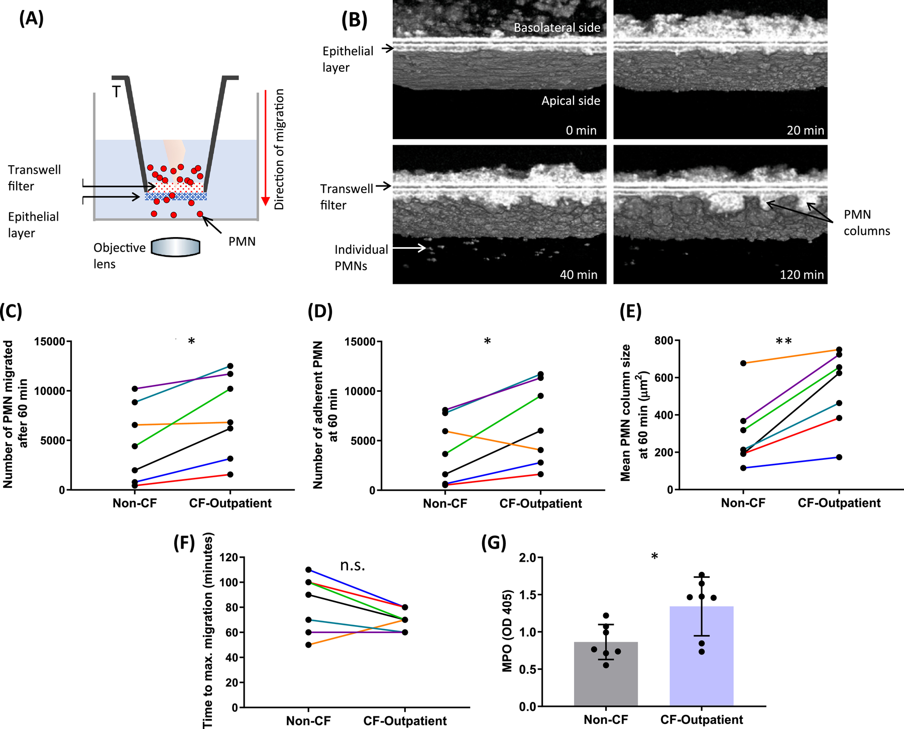
Figure 4. Neutrophil transepithelial migration across a lung epithelial monolayer.

(A) Schematic illustrating µOCT imaging of the neutrophil transepithelial migration assay. (B) Aggregation and adherence of migrated neutrophils, as well as subsequent detachment of individual neutrophils are shown in representative 3D µOCT images captured at various time points after initiation of transepithelial migration. Using the 60-minute time point to compare paired experiments, we found that there was (C) a significantly greater number of CF-outpatient neutrophils that migrated and (D) adhered to the apical side of the lung-epithelial layer. (E) En face views taken about at 10 µm below the epithelial monolayer revealed that the mean area of adherent CF-outpatient neutrophil columns was significantly larger than that of non-CF neutrophils. (F) Maximal neutrophil migration occurred more rapidly in the CF-outpatient group, and G) unstimulated, unmigrated CF-outpatient neutrophils released greater MPO (as quantified by OD405) per 500,000 neutrophils than healthy controls. N = 7 non-CF, N = 7 CF-outpatient, paired-samples on the same day. * P < 0.05; ** P < 0.01. MPO = myeloperoxidase
