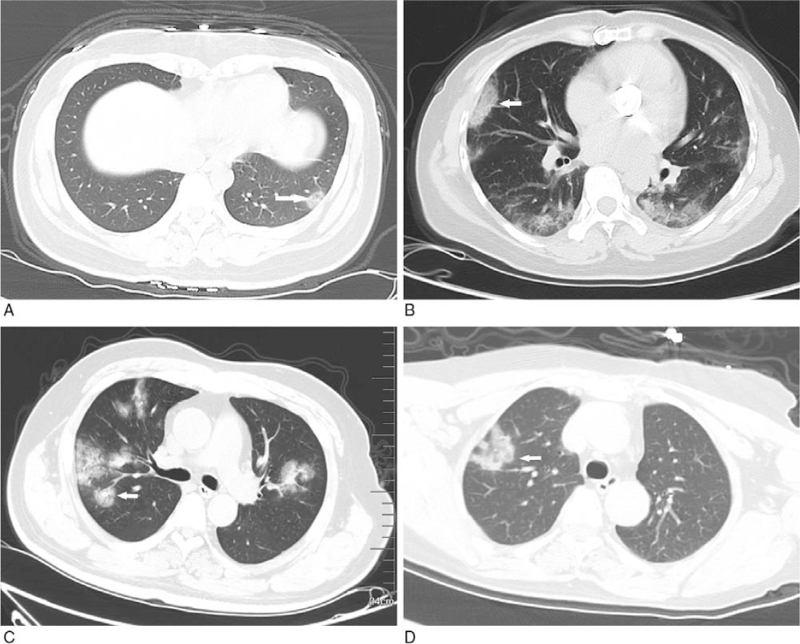
Figure 2.
GGO with clear margin. A: subpleural GGO in the lateral basal segment of the inferior lobe of left lung with uneven density (white arrow); B: bilateral pulmonary multiple GGO: subpleural GGO in the lateral segment of the middle lobe of right lung with uneven density (white arrow); C: bilateral pulmonary multiple GGO with uneven density (white arrow); D: single GGO in the superior lobe anterior segment of right lung with irregular morphology and uneven density (white arrow). GGO = ground-glass opacification.

