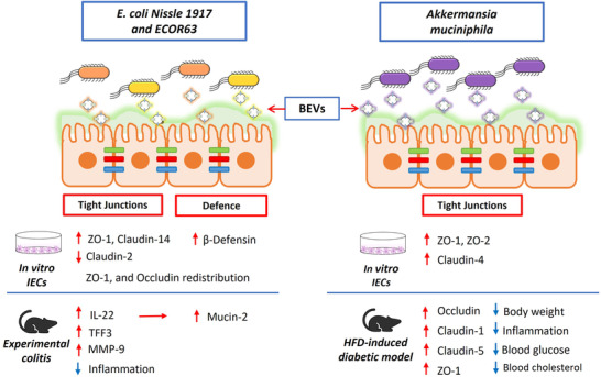
FIGURE 3.

Modulation of the gut epithelial barrier by microbiota BEVs. Schematic representation of the intestinal epithelium, where tight junction (TJ) proteins are indicated by coloured bars connecting adjacent intestinal epithelial cells (IECs). BEVs released by E. coli Nissle 1917 (EcN) and ECOR63 (left panel), or A. muciniphila (right panel) migrate through the inner mucus layer and reach the epithelium. Regulatory mechanisms influencing the integrity of the intestinal barrier known to be activated by BEVs are indicated below the drawing, specifying whether evidences were obtained from in vitro assays (culture of IECs) or in vivo models of increased intestinal permeability (experimental colitis and high fat diet (HFD)‐induced diabetic model). Upregulation/downregulation of gene transcription is indicated by red arrows. For each experimental model, the beneficial effects of BEVs counteracting disease alterations are shown by blue arrows. Overall, the regulatory effects mediated by microbiota BEVs result in gut epithelial barrier reinforcement and the subsequent reduction of intestinal permeability
