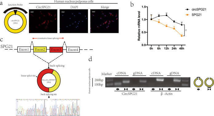
Fig. 2. Ring structure of circSPG21.
a Left, circSPG21 and probe diagram. Right, RNA-fluorescence in situ hybridization (FISH) showed that circSPG21 was mainly present in the cytoplasm (scale bar, 100 μm). The circular RNA probe was labeled with Cy-3. Nuclei were stained with 4′,6-diamidino-2-phenylindole (DAPI). b Nucleus pulposus cells were treated with 5 μg/mL actinomycin D, and the expression levels of circSPG21 and SPG21 were detected by RT-qPCR, *p < 0.05. Data are presented as the mean ± S.D., and the p values were determined by a two-tailed unpaired Student’s t-test. c Exons 2–3 of SPG21 form circSPG21. Sanger sequencing confirmed the presence of circSPG21. d Left: circSPG21 was amplified by divergent and convergent primers in cDNA and genomic DNA and separated by horizontal electrophoresis. β-actin served as a negative control. Right: a diagram of the divergent and convergent primers.

