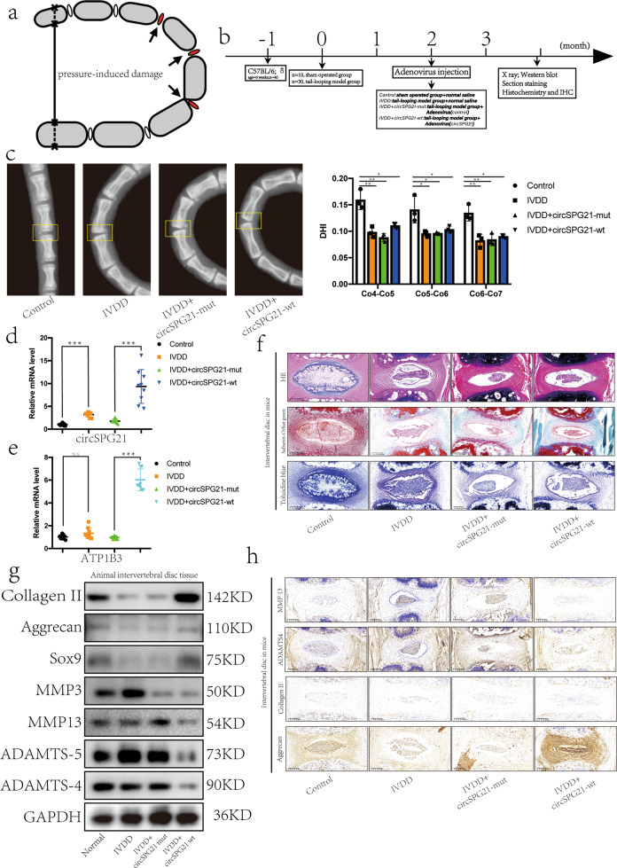
Fig. 6. Injection of circSPG21 alleviates IVDD in a mouse model.
a Schematic diagram showing the composition and principle of the tail-looping model. b Diagram showing the time sequence of in vivo experiments. c Left, the tails of mice, as observed by X-ray. Right panel, disc height index (DHI) of coccyx4–coccyx5, coccyx5–coccyx6, and coccyx7–coccyx8; n = 12, *p < 0.05, **p < 0.01. Data are presented as the mean ± S.D., and the p values were determined by a two-tailed unpaired Student’s t-test. d Expression of circSPG21 in the intervertebral discs of mice after the injection of adenovirus or saline, as detected by real-time quantitative polymerase chain reaction (RT-qPCR); n = 12, ***p < 0.001. Data are presented as the mean ± S.D., and the p values were determined by a two-tailed unpaired Student’s t-test. e Expression of ATP1B3 in the intervertebral disc of mice, as detected by RT-qPCR; n = 12, ***p < 0.001. Data are presented as the mean ± S.D., and the p values were determined by a two-tailed unpaired Student’s t-test. f Tissue sections of mice stained with hematoxylin and eosin (HE), safranin-O/fast green, and toluidine blue to show the degree of intervertebral disc degeneration (scale bar, 200 μm). g Expression of collagen II, aggrecan, SRY-box transcription factor 9 (SOX9), matrix metallopeptidase 3 (MMP3), MMP13, ADAM metallopeptidase with thrombospondin type 1 motif 4 (ADAMTS4), and ADAMTS5 in intervertebral disc tissue, as determined by western blotting. h Expression of proteins (collagen II, aggrecan, ADAMTS4, and MMP13), as determined by immunohistochemical staining (scale bar, 200 μm).

