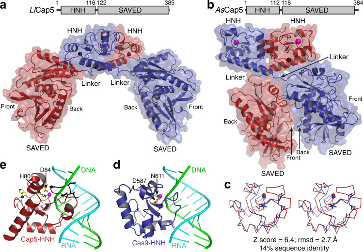
Fig. 3. Crystal structures of unliganded Cap5.
a Cartoon and surface depiction of the structure of LlCap5. Cap5 forms a homodimer, and two monomers are colored red and blue, respectively. Zn2+ ions are in the sphere and colored black. b Structure of AsCap5. Additional Mg2+ ions in the active sites of HNH domains are in sphere and colored magenta. c Stereoview of structural superposition of HNH domain from AsCap5 (red) to the one from Cas9 (blue). The structures are depicted in ribbon, and Mg2+ ions are in sphere. d Structure of Cas9-HNH domain in complex with DNA•RNA hybrid. Two residues coordinating a Mg2+ ion are in stick and colored individually, with carbon in yellow, oxygen in red, and nitrogen in blue. e Structure of AsCap5-HNH domain with the modeled DNA•RNA hybrid from d. In addition to coordination of a Mg2+ ion, a zinc finger, which is absent in Cas9-HNH domain, is also depicted in the stick, with the Zn2+ ion in black and the sulfur atom in orange. The peptide having a steric clash with the DNA strand of the modeled DNA•RNA hybrid is colored black and depicted in the stick.

