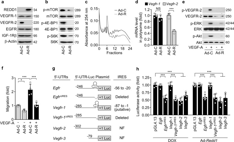
Fig. 3. REDD1 overexpression suppresses Vegfr-2 translation and angiogenesis.
a–f VEGFR-1/2 expression and VEGF-A-induced angiogenesis were examined in the HUVECs infected with control adenovirus (Ad-C) or Ad-Redd1 (Ad-R) at an MOI of 50. a, b Western blots of VEGFR-1/2, EGFR, and IGF-1Rβ (a) as well as phosphorylated mTOR, 4E-BP1, and S6K (b). c Polysome profiling using sucrose density gradient ultracentrifugation. d qRT-PCR of Vegfr-1/2 mRNAs associated with high-molecular-weight polysomes (n = 4). e Western blots of phosphorylated VEGFR-2, ERK, and Akt in the HUVECs stimulated with VEGF-A. f Quantitative migration and tube formation of HUVECs in response to VEGF-A were assessed using Boyden chamber and Matrigel-based morphogenesis assays, respectively. (n = 4). g Schematic representation of the 5′-UTRs of Egfr, Vegfr-1, Vegfr-2, and Vegfr-3; clover-like structures in the 5′-UTRs of Egfr (-54 to -25) and Vegfr-1 (−87 to −1) indicate authentic IRES and putative IRES sequences, respectively; +1 indicates the AUG start codon, and dotted lines indicate deleted regions of 5′-UTRs. NF, not found. h HEK-293 cells were cotransfected with the pGL4.13/5′-UTR-Firefly luciferase construct and Renilla luciferase pGL4.74 vector, followed by treatment with or without DOX or infection with Ad-C or Ad-Redd1. Reporter activities were measured in cell lysates using a Dual Luciferase Assay Kit (n = 5). Data are presented as the mean ± SD. *P < 0.05, ***P < 0.001; NS, not significant.

