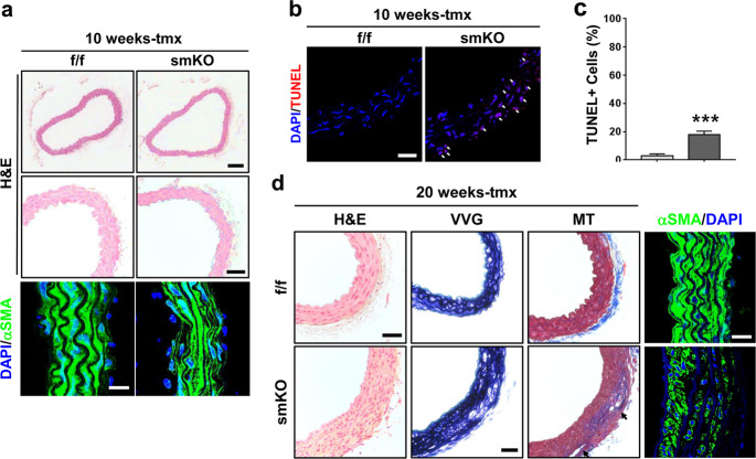
Fig. 3. Prmt1-deficient aortas exhibit VSMC apoptosis and medial degeneration.
a Histological analysis of thoracic aortas isolated from the f/f and smKO mice treated with tmx for 10 weeks. Representative images of hematoxylin and eosin (H&E) staining. Scale bar: 100 μm (upper), scale bar: 50 μm (bottom). Immunostaining for αSMA on the f/f and smKO aortas. Scale bar: 40 μm. b Representative confocal microscopic images of TUNEL-positive nuclei (red) in the f/f and smKO mouse aortas. Scale bar: 50 μm. c Quantification of TUNEL-positive nuclei in Panel b. Data represent the mean ± SD. ***P < 0.01, Student’s t-test. d Histological analysis of thoracic aortas isolated from the f/f and smKO mice treated with tmx for 20 weeks. Representative images of H&E, Verhoeff-Van Gieson (VVG), and Masson’s trichrome (MT) staining. Scale bar: 100 μm. Immunostaining for αSMA in the f/f and smKO aortas. Scale bar: 40 μm.

