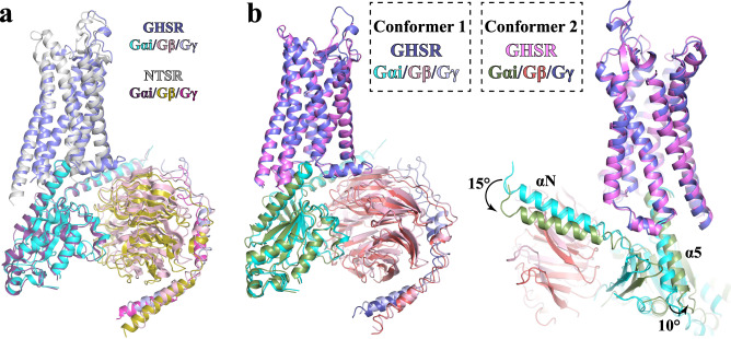
Fig. 6. Structures of the ghrelin-GHSR-Gi complex in Conformer 1 and 2.
a Structural alignment of the ghrelin-GHSR-Gi complex in Conformer 1 with the neurotensin-NTSR-Gi complex in the canonical state (PDB ID 6OS9). GHSR and NTSR are colored in slate and light gray, respectively. Gαi, Gβ, and Gγ subunits are colored in cyan, pink, and light blue, respectively, in the structure with GHSR, and in dark purple, olive, and warm pink, respectively, in the structure with NTSR. The Gi-coupling mode is highly similar. b Structural alignment of the ghrelin-GHSR-Gi complex in Conformer 1 and 2. Gαi, Gβ, and Gγ subunits are colored in cyan, pink and light blue, respectively, in Conformer 1, and in forest, ruby, and deep blue, respectively, in Conformer 2. GHSR is colored in slate in Conformer 1 and violet in Conformer 2.

