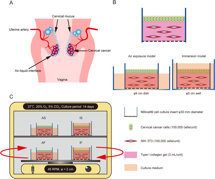
Fig. 1.
Schematic illustration of the specific microenvironments of cervical cancer and the culture model. (A) Schematic illustration of the specific microenvironments of cervical cancer. (B) Illustrations showing the collagen gel culture model and the double-dish air-liquid interface culture method. To replicate the air-liquid interface, the culture fluid level of the outer dish was adjusted to be at the height of the collagen gel in the inner dish. Cervical cancer cells were seeded on a collagen gel embedded with NIH 3T3 cells or a collagen gel without NIH 3T3 cells (control). (C) To generate fluid flow, culture dishes were placed on a rotatory shaker in a CO2 incubator. The cells were cultured under 4 conditions, namely immersion under static flow (IS), immersion under dynamic flow (IF), air exposure under static flow (AS) and air exposure under dynamic flow (AF).

