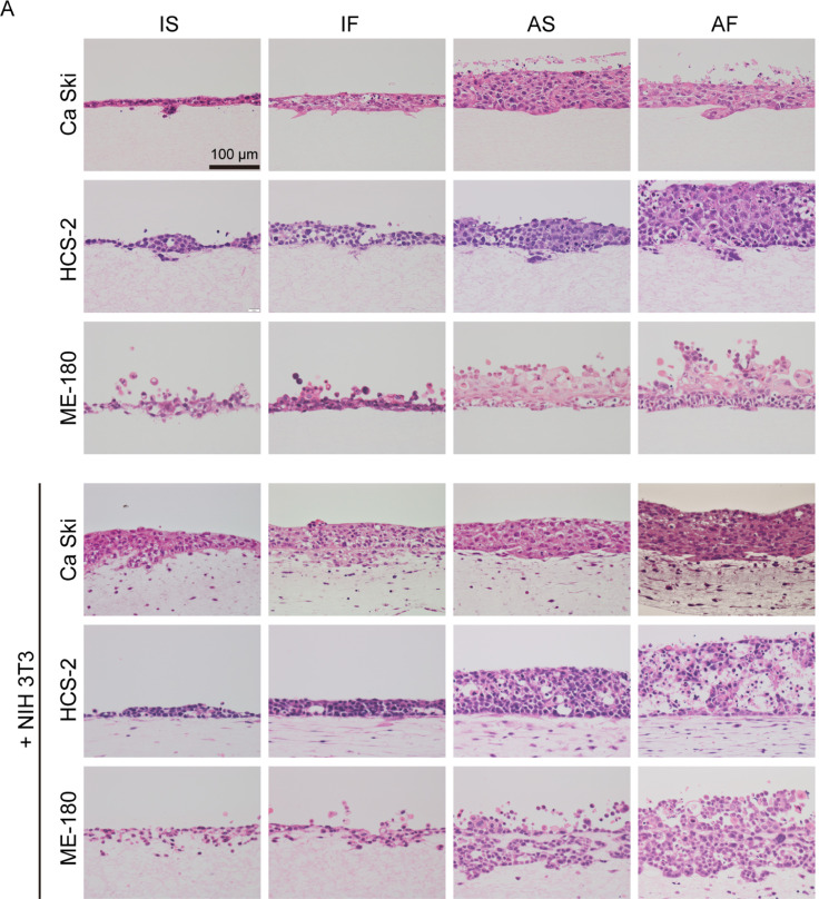
Fig. 2.
Effects of fluid flow and air exposure on the cellular kinetics of cervical cancer cells. (A) Representative images at day 14. Under IS conditions, Ca Ski, HCS-2 and ME-180 cells cultured without NIH 3T3 cells had a flat cytoplasm, formed a thin layered structure and invaded the collagen matrix at only a few locations. Fluid flow promoted cytoplasmic hypertrophy and thickening of the cellular layer of monocultured Ca Ski, HCS-2 and ME-180 cells. Fluid flow and air exposure synergistically induced cellular hypertrophy and a thickening of the cellular layer of Ca Ski, ME-180 and HCS-2 cells. ME-180 and HCS-2 cells, but not Ca Ski cells, had a thicker cellular layer under AF conditions than under AS conditions. Under IS conditions, NIH 3T3 cells promoted cellular hypertrophy and increased the cellular layer thickness of Ca Ski cells but not the other cell types. Fluid flow increased the cellular layer thickness of HCS-2 cells co-cultured with NIH 3T3 cells, but this effect was not observed in Ca Ski and ME-180 cells. Fluid flow increased the number of invasive spots for Ca Ski cells co-cultured with NIH 3T3 cells. Under static flow conditions, air exposure increased the cellular layer thickness of all 3 cervical cancer cell types. Fluid flow and air exposure significantly increased the cellular layer thickness of Ca Ski and ME-180 cells. By contrast, HCS-2 cells co-cultured with NIH 3T3 cells exhibited less thickening of the cellular layers and a smaller degenerative area under AF conditions. Bar = 100 μm. (B) The thickness of the cellular layers. *P < 0.05. **P < 0.001. Abbreviations: AF, air exposure under dynamic flow; AS, air exposure under static flow; IF, immersion under dynamic flow; IS, immersion under static flow. Data are shown as the mean ± standard deviation of 3 measurements.

