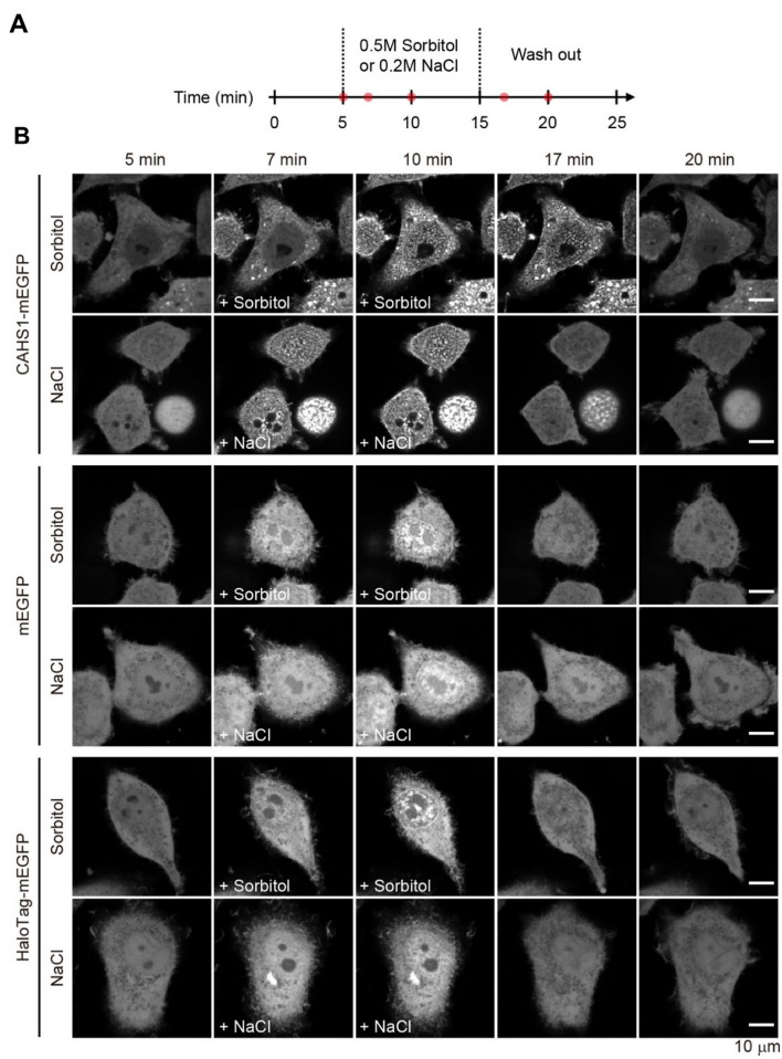
Figure 3.
Real-time monitoring of the reversible formation of CAHS1 protein particles. (A) Timeline of time-laps imaging with hyperosmotic shock. The red dots represent the time points when the representative cells are presented in panel (B). (B) Time-laps images of HeLa cells overexpressing the CAHS1-mEGFP, mEGFP, and Halo Tag-mEGFP proteins. A sorbitol or sodium chloride solution was added at 5 min. After 10 min, the cells were washed with a flesh medium using a microfluidics system.

