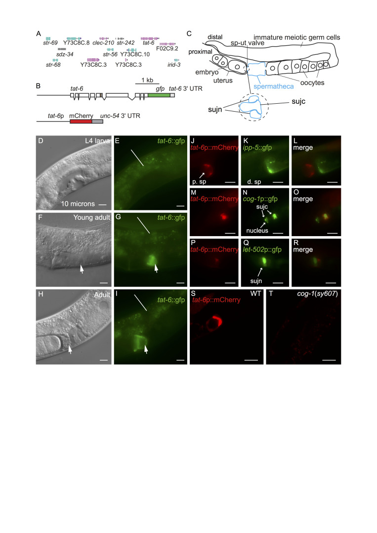
Figure 1. tat-6 is expressed in sujc cells.
A. The tat-6 chromosomal region. The genes shown are those present in the fosmid used to generate the tat-6::gfp reporter shown in B. B. Schematics of the tat-6 reporters used in this study. Boxes represent exons. The upper construct encodes a TAT-6::GFP fusion protein in which GFP is fused to the whole of TAT-6. The beige-colored box indicates the part of TAT-6 containing the P-type ATPase motif. The reporter gene was generated by inserting gfp-encoding sequences into the fosmid (Sarov et al. 2012). The fosmid also contains the genes (shown in A) to the left and right of tat-6 in the C. elegans genome as well as intragenic sequences (Sarov et al. 2012). The tat-6::gfp reporter has the tat-6 3ʹ untranslated region (UTR). The lower construct is a transcriptional reporter in which mCherry expression is driven by a 1.4 Kb fragment from the tat-6 promoter. This construct contains the 3ʹ-UTR of the unc-54 gene. C. A schematic showing one arm of the gonad in an adult hermaphrodite. The spermatheca is highlighted in blue. ‘sp-ut valve’ indicates the position of the spermathecal-uterine valve. The expanded region shows a schematic cross section of the valve as it exists prior to ovulation. In such animals, the sujc syncytium occupies the core of the valve and is surrounded by a toroidal syncytium, sujn (Kimble and Hirsh 1979). During the first ovulation, sujc is displaced from the core of the valve. D-I, micrographs of the mid-body regions of hermaphrodite worms harboring the tat-6::GFP transgene shown in B. In D, F and H the worms were viewed with Nomarksi differential interference contrast (DIC) optics; E, G and I show the same worms viewed with fluorescence optics. In F and H the arrows indicate the position of the spermathecal-uterine valve. The uterus is to the left of the arrows, the spermatheca to the right. In G and I the arrows indicate GFP fluorescence in the gonad. The lines in E, G and I indicate background autofluorescence from the intestine. H shows a hermaphrodite that contained fertilized eggs in the uterus; the egg in closest proximity to the spermatheca is partially enveloped by material containing the TAT-6::GFP fusion protein. J,K,L. Fluorescent micrographs of an adult hermaphrodite carrying tat-6p::mCherry and ipp-5::GFP transgenes. The worms were mounted for photography so that the uterus was to the left. The ipp-5::GFP transgene is expressed in distal spermathecal cells (d. sp) at the junction with the ovary (Bui and Sternberg 2002). Note the lack of overlap between the mCherry and GFP fluorescence signals. M,N,O. Fluorescence micrographs of a hermaphrodite harboring tat-6p::mCherry and cog-1p::GFP transgenes. The cog-1 transgene is expressed in the sujc syncytium, which forms the core of the spermathecal-uterine valve (Palmer et al. 2002). To the right in the panels, the mCherry and GFP signals overlap. Note that while the distal-most part of the sujc syncytium is firmly within the center of the core, the sujc cell nuclei protrude into the uterus and are almost 10 microns from the distal part of the cell (Palmer et al. 2002). While some GFP encoded by the cog-1p::GFP transgene is nuclear, the mCherry signal is strongest in the distal part of the syncytium. P,Q,R. Fluorescent micrographs of a hermaphrodite harboring tat-6p::mCherry and let-502p::GFP transgenes. The let-502p::GFP transgene is strongly expressed in sujn cells (Wissmann et al. 1999). S,T. Confocal fluorescence micrographs of adult hermaphrodites harboring the tat-6p::mCherry transgene. S shows an otherwise wild-type hermaphrodite; T shows a cog-1 mutant. Scale bars in all panels indicate 10 microns.

