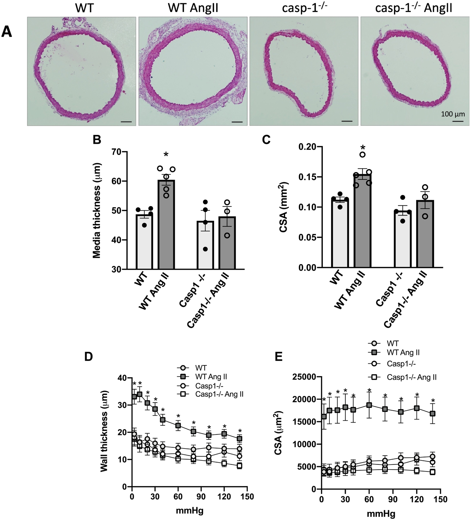
Figure 4. Ang-II treatment induces vascular remodeling via inflammasome.

Representative (A) and quantitative histomorphometric analysis of aorta media thickness (B) and aorta cross-sectional area (C). Wall thickness (D) and cross-sectional area (E), determined in a myograph for pressurized arteries, in mesenteric resistance arteries. Aorta and mesenteric arteries from Ctrl (WT) and Casp1−/− mice treated with Ang-II (490 ng/min/kg for 14 days with ALZET osmotic minipumps). Mesenteric arteries were gradually pressurized in passive conditions (0Ca2+ Krebs Henseleit Buffer). Values are reported as mean ± s.e.m. Black lines on the representative images from aortic CSA represent 100 μm. N = 3 to 5. Values are reported as mean ± s.e.m. *P <0.05, vs. WT
