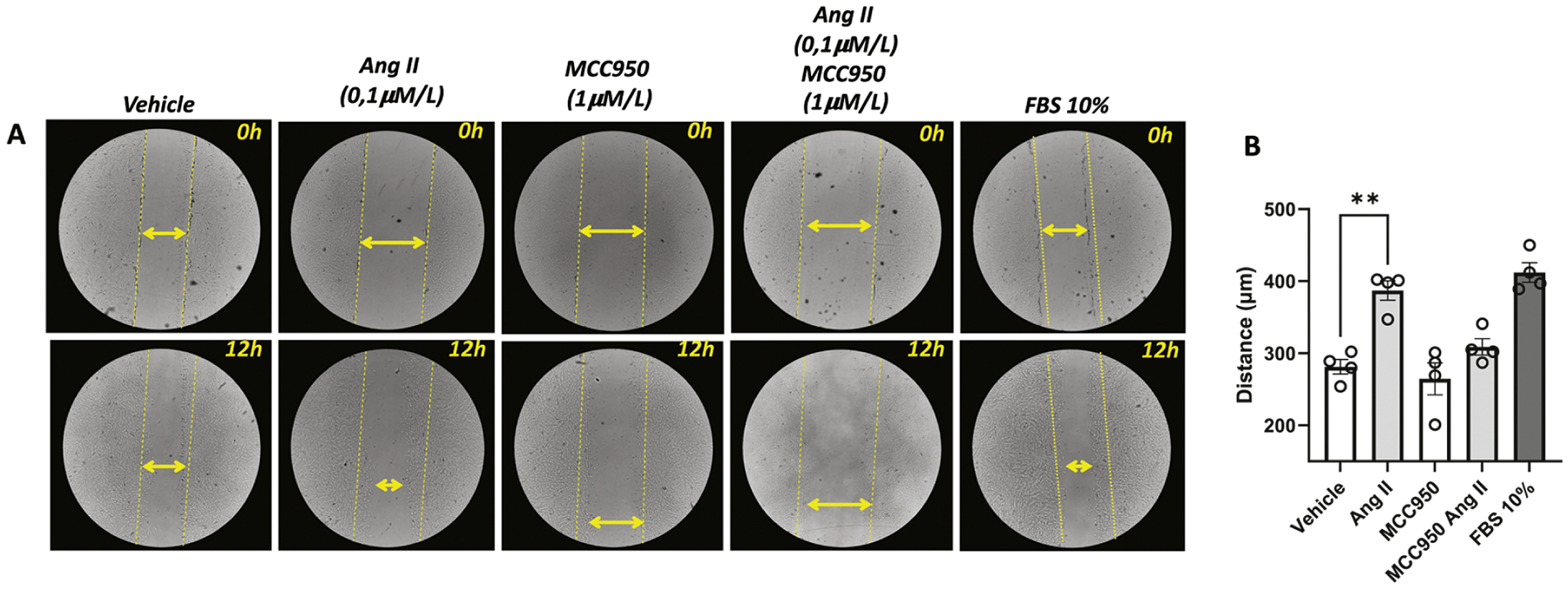
Figure 7. Angiotensin-II induces vascular migration via inflammasome.

Cell migration was analyzed by scratch wounding assay. RASMC were counted using (1×10⁵ cells per well) and seeded in 12-well dark plates. Photos were obtained right after the scratch and 12 h after incubation with Ang-II (0,1 μM). Some experiments were performed in the presence of a selective NLRP3 antagonist (MMC950, 1 μM, 30 min). Fetal Bovine Serum (FBS, 10%) was used as a positive control. Images represent individual experiments from the same well. Yellow lines represent the distance of the scratches before any stimulus. Yellow double arrows represent the distance of the scratches after 24h of the stimulus. N= 4. Values are reported as mean ± s.e.m. **P<0.05 vs. vehicle.
