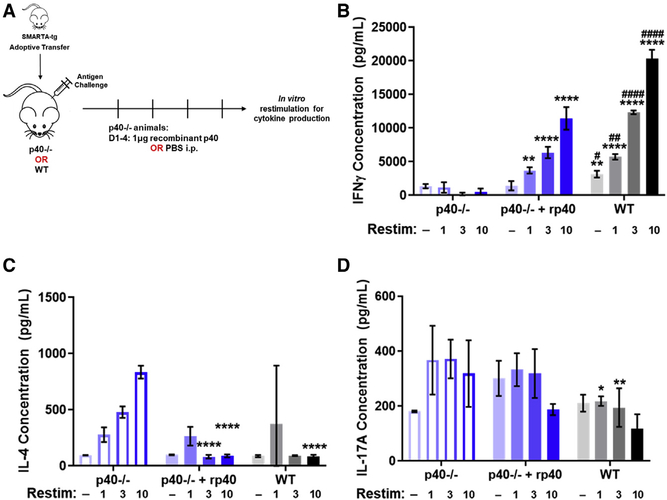
Figure 2. Administration of recombinant p40 allows for antigen-specific T cell differentiation.
(A) Schematic of experimental design. SMARTA CD4+ T cells from CD45.1+ donors were sorted and transferred into 45.2+ p40−/− animals or WT animals and challenged 24 h later with GP61–80 and LPS. p40−/− recipients were divided into two groups, one that received 1 mg of recombinant p40 and the other that received sterile PBS from days 1 to 5 after transfer. SMARTA T cells were harvested from these animals at day 5 and restimulated in vitro to analyze for T cell differentiation by cytokine production.
(B–D) Splenocytes harvested 5 days after challenge were normalized to SMARTA T cell number and restimulated in vitro with GP61–80 for 48 h. Restimulation doses are in mg. Concentration of (B) IFNγ, (C) IL-4, and (D) IL-17A in supernatant from splenocytes of SMARTA T cells transferred into p40−/− animals treated with PBS (left, blue open bars), p40−/− animals given 1 μg of recombinant p40 (middle, blue filled bars), or WT animals (right, gray filled bars) (n = 4).
Data in this figure are representative of one independent experiment where n refers to the number of biological replicates. (B)–(D) are displayed as mean ± SEM. *,#p < 0.05, **,##p < 0.01, ***,###p < 0.001, ****,####p < 0.0001: *significance relative to the p40−/− PBS-treated animals; #significance relative to the p40−/− animals, which received recombinant p40. Anything unmarked is considered not significant.

