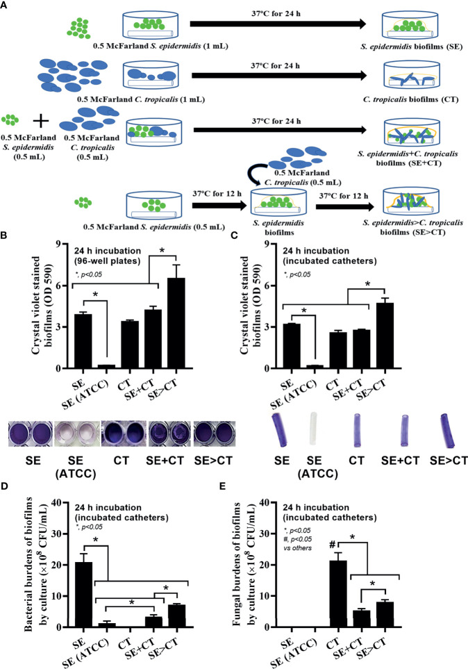
Figure 1.
Schema of the in vitro experiments for the preparation of biofilms from Staphylococcus epidermidis (SE), Candida tropicalis (CT), SE simultaneously mixed with CT (SE + CT), and preformed SE biofilms following CT (SE > CT) (A) are demonstrated. Characteristics of biofilms from SE, either the clinically isolated strain (SE) or the standard strain [SE (ATCC)], CT, SE + CT, and SE > CT as determined by intensity of crystal violet color on 96-well plates and incubated catheters with the representative pictures (B, C); bacterial and fungal burdens on catheter biofilms (D, E) are demonstrated (independent triplicate experiments were performed). The results were from three independent experiments in triplicate as the mean ± SEM; p < 0.05 was considered statistically significant. *p < 0.05 was considered statistically significant in the same group whereas #p < 0.05 was those in the other groups.

