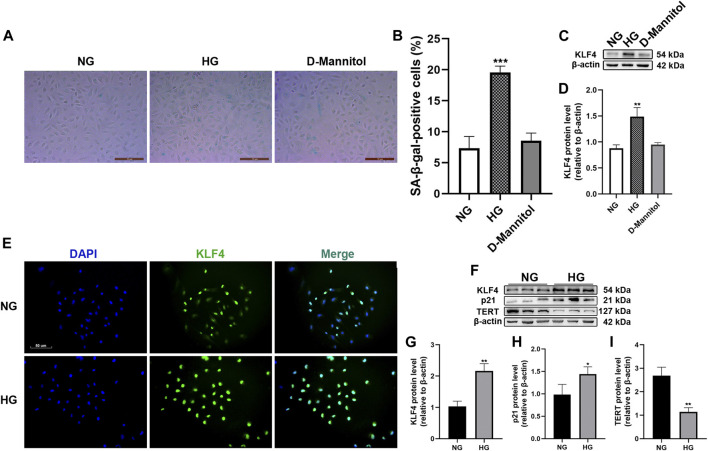
FIGURE 1.
Effect of HG exposure on KLF4, p21, and TERT expression in HUVECs. (A) For the studies comparing the effects of mannitol, HUVECs were incubated with media consisting of NG or HG, and D-mannitol for 48 h. Cells were fixed and stained for SA-β-gal activity. The images are taken by ×10magnification. (B) Histogram represents the percentage of SA-β-gal-positive cells per microscopic field. (C) Cell lysates were used to determine the KLF4 protein levels. (D) Results were normalized to controls, and histograms represent the relative intensity of KLF4. Values represent mean ± SEM (n = 3–4 per group). **p < 0.01, ***p < 0.001, significantly different from NG. (E) HUVECs were also cultured either in NG or HG media for 48 h, and intracellular KLF4 levels were measured by immunofluorescence staining. The images were taken at ×100 magnification and sections were stained with KLF4 (green) and DAPI (blue). A representative image from three separate experiments is illustrated. (F) KLF4, p21, and TERT protein levels were determined by immunoblotting. (G–I) Results were normalized to controls, and histograms represent the relative intensity of KLF4, p21, and TERT. Values represent mean ± SEM (n = 3–4 per group). **p < 0.01, ***p < 0.001, significantly different from NG.

