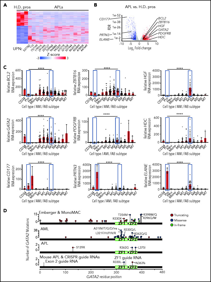
Figure 1.
GATA2 expression in normal and malignant hematopoietic cells. (A) Heat map of the 4094 DEGs between primary human APLs and healthy donor promyelocytes (H.D. pros) by RNA-seq fold change ≥2 and FDR <0.05. (B) Volcano plot of the samples from panel A. Expression changes of BCL2, ZBTB16, HGF, GATA2, PDGFRB, HDC, CD177, PRTN3, and ELANE are labeled. (C) BCL2, ZBTB16, HGF, GATA2, PDGFRB, HDC, CD177, PRTN3, and ELANE expression in flow purified healthy donor human CD34+ progenitors (CD34), promyelocytes (Pro), neutrophils (Neu), monocytes (Mono), and the AML French-American-British subtypes M0-M7 by RNA-seq using the AML TCGA data set.6 Promyelocytes and M3 (APL) subtype samples are highlighted in blue. Statistical significance was determined by edgeR, which includes correction for multiple testing. P values are corrected for multiple testing. (D) Distribution of GATA2 mutations from patients with Emberger syndrome, monocytopenia and mycobacterial infection (MonoMAC) syndrome, AML, or APL,1-5,7,11,30,34,59-64 as well as those from mouse APLs identified by exome sequencing. The locations of CRISPR guide RNAs used in this study (labeled exon 2 and ZF1) are annotated with dashed lines. ****P < .0001. UPN, unique patient number; ZF1, zinc finger domain 1; ZF2, zinc finger domain 2.

