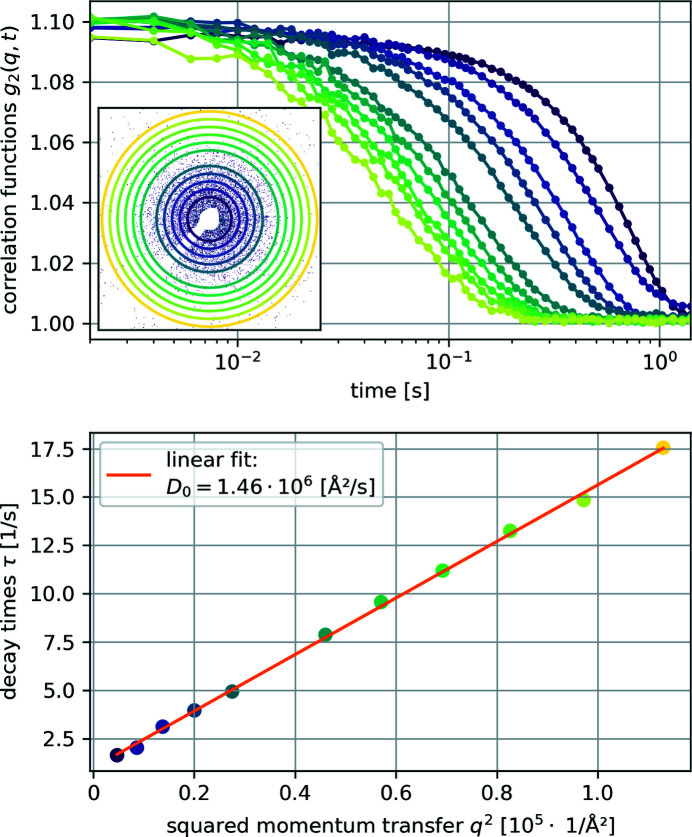
Figure 4.
Top: calculated intensity correlation functions g 2(q, t) from the measurements on silica-coated hematite ellipsoids dispersed in glycerol. The inset shows one of the recorded scattering patterns and the rings mark the q used for the other plots. Bottom: fitted decay times τ plotted against q 2 showing a linear decency and the linear fit to estimate the free particle diffusion coefficient D 0. The color coding of each data point marks the corresponding intensity correlation function g 2(q, t) and the ring/radius in the inset.

