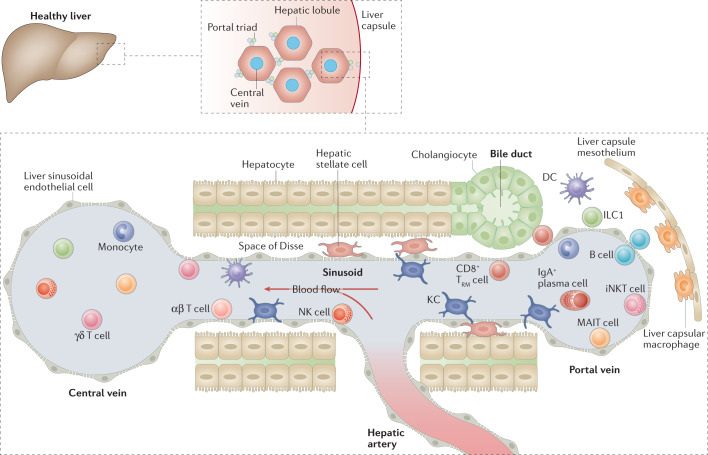
Fig. 2. Immune landscape of the healthy liver.
The liver is composed of lobules that are repeated hexagonal anatomical units. These lobules consist of hepatocytes organized around a central vein that is connected to the hepatic artery and the portal vein via a sinusoidal network. The bile produced by hepatocytes is released into the gastrointestinal tract via bile ducts composed of cholangiocytes. The hepatic artery, portal vein and bile duct together form the portal triads found between hepatic lobules. The blood flow along the portal to central axis (from portal vein and hepatic artery to central vein) creates gradients of oxygen and nutrients, resulting in a spatial division of labour among hepatocytes, a phenomenon known as metabolic zonation. A large spectrum of immune cells is found in the liver. Some circulate or temporarily patrol the hepatic sinusoids or the liver parenchyma, such as natural killer (NK) cells, γδ T cells, CD4+ and CD8+ αβ T cells, monocytes, B cells, invariant NKT (iNKT) cells, mucosal-associated invariant T (MAIT) cells and dendritic cells (DCs). Long-lived resident cells, such as Kupffer cells (KCs), CD8+ tissue-resident memory T (TRM) cells and type 1 innate lymphoid cells (ILC1s) are also found. Liver lobules also have a spatially polarized immune system, known as immune zonation, with KCs, iNKT cells, CD8+ TRM cells and IgA+ plasma cells being particularly enriched in the periportal regions. KCs, which specifically reside in the sinusoids in close contact with liver sinusoidal endothelial cells, are the most abundant immune cell population in the liver. They also establish connections with hepatic stellate cells and hepatocytes in the space of Disse. Another resident macrophage population occupies the liver capsule and these cells are known as liver capsular macrophages.

