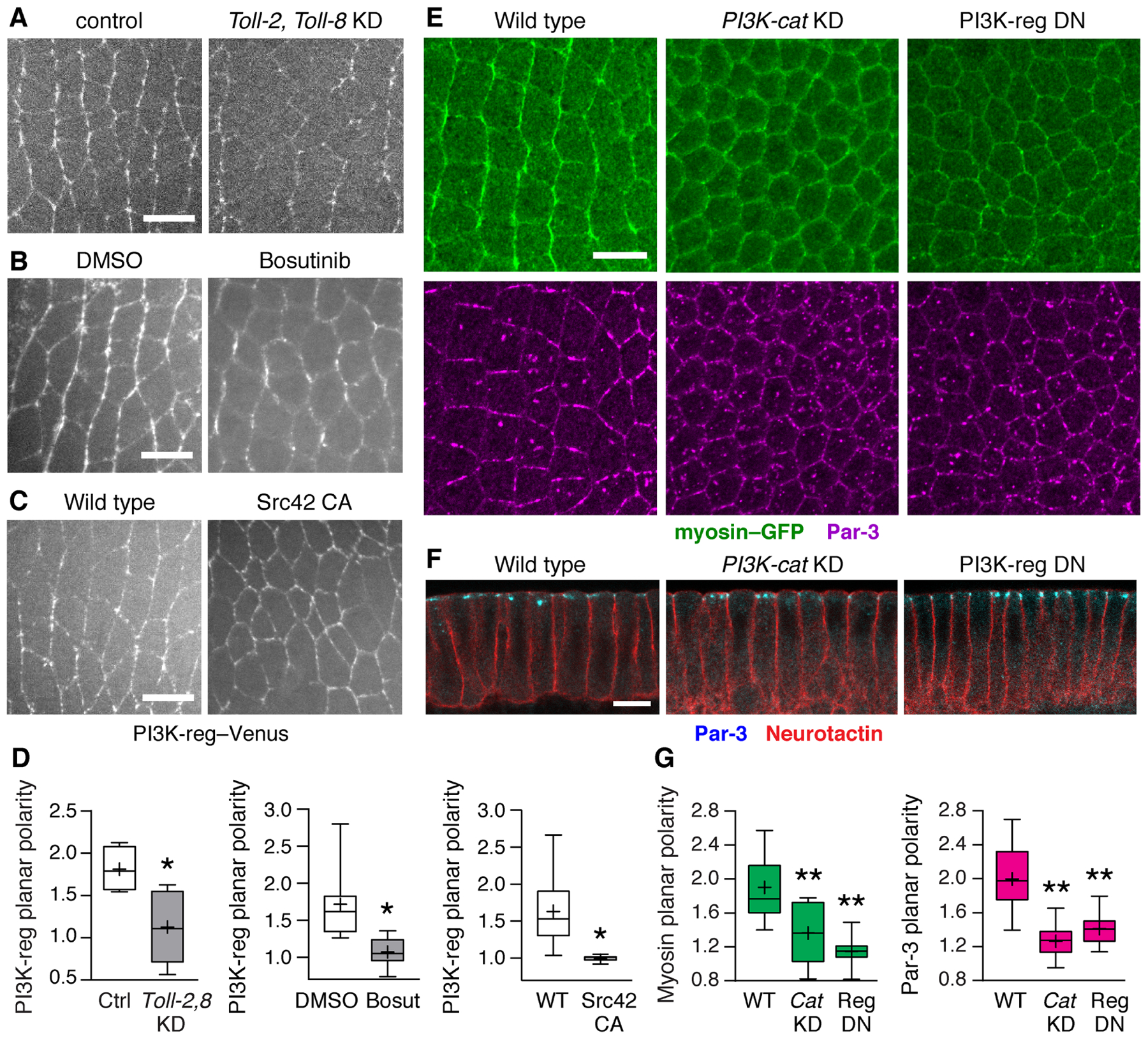
Figure 4. The PI3K complex is necessary for planar polarity during convergent extension.

(A–C) PI3K-reg–Venus localization in (A) embryos injected with dsRNAs to Toll-2 and Toll-8 (Toll-2,Toll-8 KD) and control embryos injected with Toll-3 dsRNA, (B) embryos injected with Bosutinib (100 μM Bosutinib in 0.6% DMSO) and DMSO controls (0.6% DMSO) (final concentrations), and (C) embryos expressing constitutively active Src42 (Src42 CA) and wild-type Gal4 controls. (D) PI3K-reg–Venus planar polarity (vertical-to-horizontal edge intensity ratio). (E) Localization of myosin II (myosin–GFP, top panels) and Par-3 (bottom panels) in wild-type (WT), PI3K-cat KD, and PI3K-reg DN embryos. (F) Apical-basal polarity occurs normally in PI3K-cat KD and PI3K-reg DN embryos. (G) Myosin–GFP planar polarity (vertical-to-horizontal edge intensity ratio) and Par-3 planar polarity (horizontal-to-vertical edge intensity ratio). Boxes, 2nd and 3rd quartiles; whiskers, min to max; horizontal line, median; +, mean. Living stage 7 embryos are shown in (A–C), fixed stage 7 embryos in (E and F), 3–14 embryos/genotype. *p<0.03, **p<0.001, Welch’s t-test. Anterior left, ventral down (A, B, C, and E). Cross sections, apical up (F). Bars, 10 μm. See also Figures S5 and S6.
