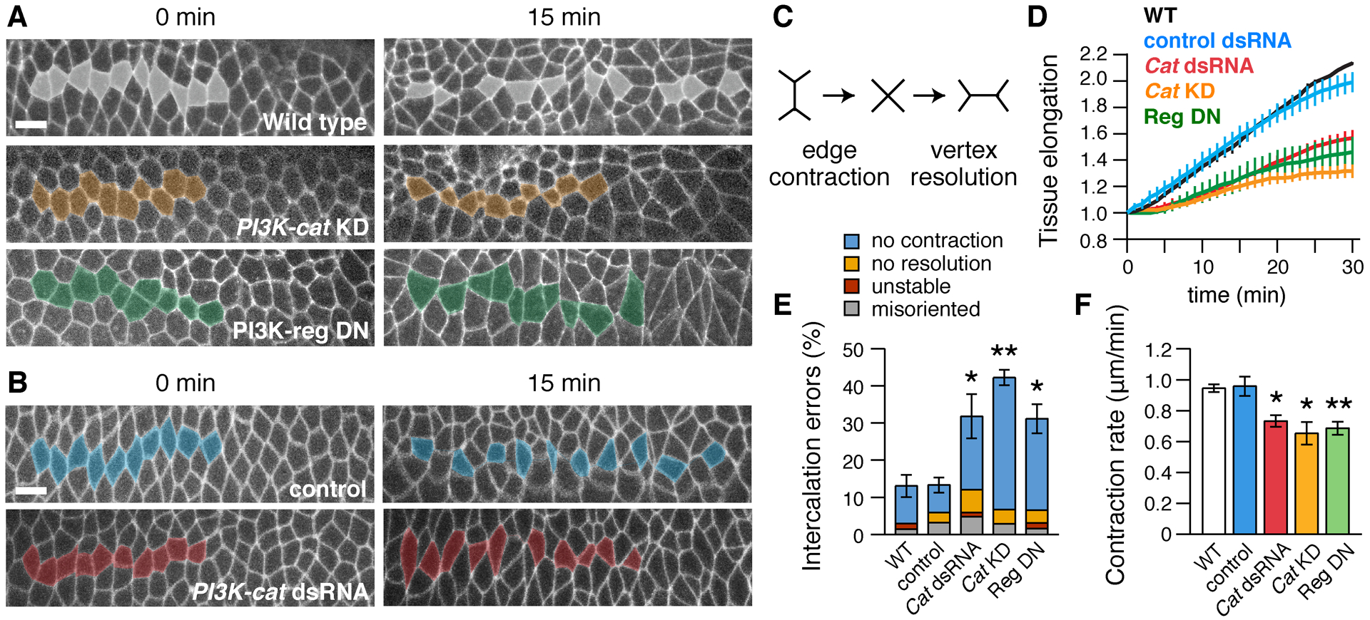
Figure 5. PI3K is required for cell intercalation and convergent extension.

(A,B) Stills from movies of (A) wild-type (WT), PI3K-cat KD, and PI3K-reg DN embryos expressing Spider–GFP and (B) control (Toll-3 dsRNA-injected) and PI3K-cat dsRNA-injected embryos expressing Resille–GFP (0 min, onset of elongation). Anterior and posterior cells become separated by intercalation in WT, but many cells fail to separate in PI3K-defective embryos. (C) Schematic of cell intercalation. (D) Axis elongation (tissue AP length relative to t=0) is reduced in PI3K-cat KD (p=0.003 at t=30 min), PI3K-reg DN (p=0.03), and PI3K-cat dsRNA embryos (p=0.01). (E) Intercalation errors are increased in PI3K-cat KD, PI3K-reg DN, and PI3K-cat dsRNA embryos. (F) The rate of vertical edge contraction is reduced in PI3K-cat KD, PI3K-reg DN, and PI3K-cat dsRNA embryos. (D–F) Mean±SEM, 3–5 living stage 7–8 embryos/genotype. *p<0.05, **p≤0.01, Welch’s t-test. Anterior left, ventral down. Bars, 10 μm.
