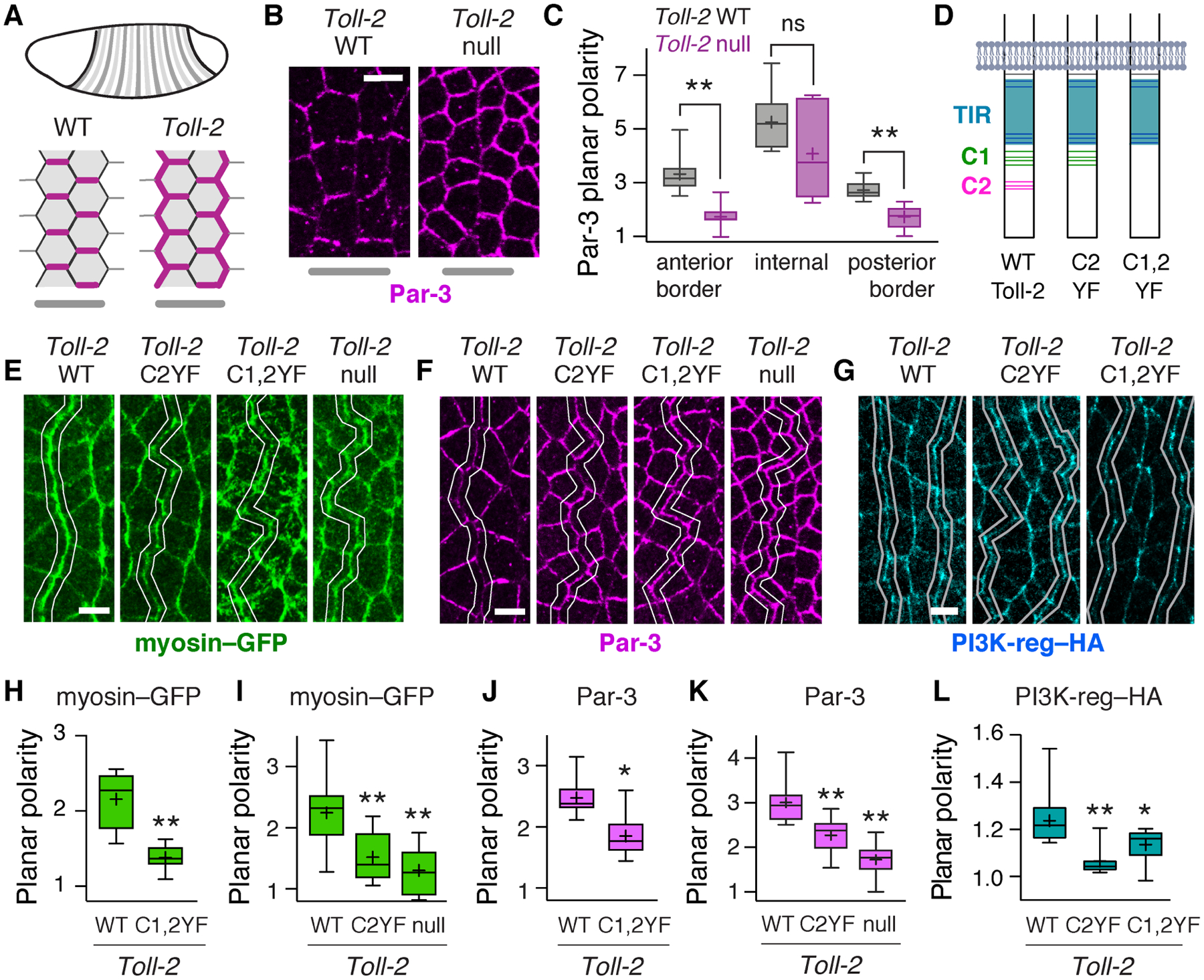
Figure 6. Toll-2 tyrosine phosphorylation is required for planar polarity.

(A) Toll-2 is required for planar polarity at Toll-2 stripe borders. Par-3 (magenta), Toll-2 stripes (gray). (B,C) Par-3 planar polarity is defective at Toll-2 stripe borders in Toll-2 null mutants, identified by Wingless staining. (D) Toll-2 cytoplasmic domain. Gray, membrane. Blue, TIR domain. Green and magenta, C-terminal tyrosine clusters C1 and C2. Tyrosines, horizontal lines. (E–G) Localization of myosin–GFP (E), Par-3 (F), and PI3K-reg–HA (G) at Toll-2 stripe borders (white lines) identified by Wingless staining in embryos expressing the indicated Toll-2 variants from the Toll-2 locus. (H–L) Myosin–GFP planar polarity (vertical-to-horizontal edge intensity ratio) (H,I), Par-3 planar polarity (horizontal-to-vertical edge intensity ratio) (J,K), and PI3K–HA planar polarity (vertical-to-horizontal edge intensity ratio) (L) at Toll-2 stripe borders. Toll-2C1,2YF images in E and F are from the same embryo. Toll-2 null mutant images in B and F are from the same embryo. Boxes, 2nd and 3rd quartiles; whiskers, min to max; horizontal line, median; +, mean, 6–16 fixed stage 7 embryos/genotype, *p<0.03, **p<0.01, Welch’s t-test. Anterior left, ventral down. Bars, 5 μm. See also Figure S6.
