Figure 7.
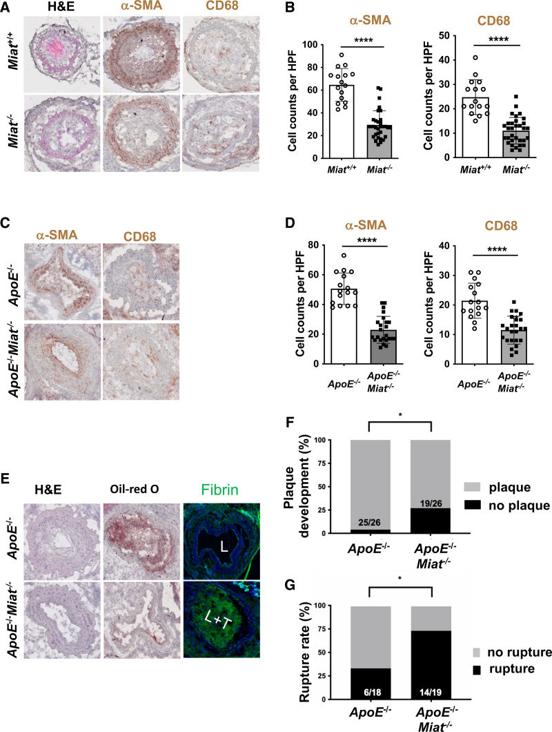
MIAT deletion affects smooth muscle cell proliferation and plaque vulnerability in vivo. A, Morphology (H&E), immunostaining for CD68 and smooth muscle cell α-actin (αSMA) in Miat–/– mice versus littermate Miat wildtype controls on carotid ligation injury. B, Analysis of cell counts (8 times/animal) per high-power field (HPF) for αSMA and CD68 from A. C, Morphology (H&E), immunostaining for CD68 and αSMA in ApoE–/–Miat–/– and ApoE–/– (Miat wildtype) controls on exposure to the inducible plaque rupture model (incomplete ligation and cuff placement). D, Cell counts (8 times/animal) per HPF for αSMA and CD68 from C. E, H&E, Oil-Red O (indicating lipid deposition), and cross-linked immunofluorescent fibrin staining (indicating an atherothrombotic event) in ApoE–/–Miat–/– versus Apo–/– Miat+/+ littermate controls (L indicates lumen; L+T, lumen with thrombus). F, Plaque development (in %) when using the inducible plaque rupture model in ApoE–/–-Miat–/– versus ApoE–/–Miat+/+ mice. G, Rupture ratio (in %) in the inducible plaque rupture model comparing ApoE–/–Miat–/– versus ApoE–/– (Miat wildtype) controls. Data were analyzed by Student t test (B, D). Fisher exact test was used to determine plaque development and rupture ratio. *P<0.05; **P<0.01. H&E indicates hematoxylin and eosin.

