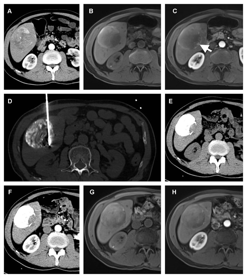
Figure 4.
A 65-year-old male with a solitary HCC lesion in the right lobe of the liver. The lesion is 75 mm in size, and TACE is performed (A). After TACE, the MR imaging shows residual enhancement in the edge of the tumor (white arrow) (B and C). A percutaneous MWA is performed in the residual enhancement area (D). After one month of ablation, no residual tumor is detected (E and F). Four months after ablation, the patient is still within the Milan Criteria (G and H), which indicates a successful downstaging case.

