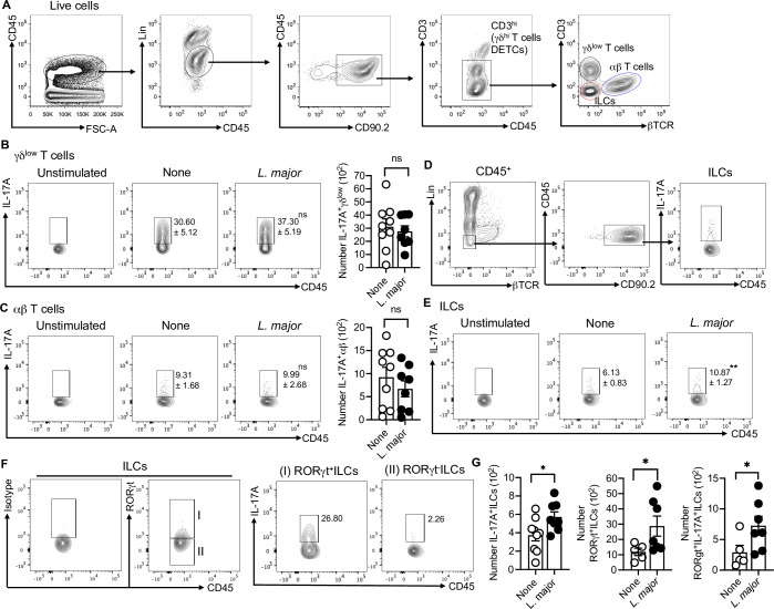
Fig 1. L. major infected skin contains IL-17A-producing RORγt+ILCs.
(A) Gating strategy to identify skin T cells and ILCs. (B) Percent and number of IL-17Α+γδlow T cells in the skin of uninfected (None) and L. major infected mice at week one. (C) Percent and number of IL-17Α+αβ T cells in the skin of uninfected (None) and L. major infected mice at week one. (D) Together with A, gating strategy to identify IL-17A+ILCs in skin. (E) Percent of IL-17Α+ILCs in the skin of uninfected (None) and L. major infected mice at week one. (F) Gating strategy to identify RORγt+IL-17A+ILCs in skin at week one. (G) Number of IL-17Α+ILCs, RORγt+ILCs and RORγt+IL-17A+ILCs in skin of uninfected (None) and L. major infected mice at week one. For intracellular staining cells were stimulated with PMA/Ion for 4 hours. Unstimulated cells were used as control to define the gates (B,C,E). Data are from two experiments with a total of five to nine mice in each group (B,C,E,G). Number within the flow plot show percent of IL-17A+ cells with SEM (B,C,E). Error bars shows SEM. Two-tailed unpaired Student’s t-test with Welch’s correction. *p<0.05, **p<0.01, ***p<0.001.

